Search Submit
Close

Radiology and Imaging (RI)

DOI:10.24983/scitemed.ri
Online ISSN: Coming soon

Radiology and Imaging (RI)

Recognized as an open access and peer reviewed journal, RI has the objectives to enhance diagnostic, therapeutic and preventive modalities and the talents of physicians, as well as to manage clinical practice in radiology. RI plays a leading role to fill the acute gap in the science and practice of medical image and allied disciplines by providing an international forum for sharing the latest discoveries and up-to-date reviews.

RI promotes submission of articles on variety of disciplines, which include medical imaging, radiotherapy, pediatric radiology, musculoskeletal radiology, oral and maxillofacial radiology, chest radiology, abdominal radiology, cardiovascular radiology, diagnostic radiology, surgical radiology, molecular imaging, breast imaging, and medical imaging technologies like ultrasound, x-ray, computed tomography, magnetic resonance imaging, and positron emission tomography.

RI incorporates manuscripts in the forms of original article, review article, short communications, case reports, image, and editorial comment. It also provides a great deal of encouragement to authors to contribute articles on the basis of its originality, importance, interdisciplinary interest, and most importantly, outstanding quality. All above mentioned submissions to the RI are subject to impartial and thorough peer-reviewed process.
Coming Soon...
Coming Soon...

    Coming Soon...

Editorial Board

  • José Antonio Franchini Ramires, MD, PhD

    Head Professor, Department of Cardiology
    University of São Paulo
    Brazil (Brasil)
    Jose Antonio F. Ramires, MD, PhD has completed his PhD at the age of 28 years from University of São Paulo Medical School, Brasil and postdoctoral studies from the same University. He was the Scientific Director, Learn Director and General Director of the Instituto do Coração-INCOR (Heart Institute) of the University of São Paulo. Currently, he is Head Professor of Cardiology of the Instituto do Coração-INCOR and coordinator of medical graduate of the INCOR. Dr. Ramires was President of the São Paulo State Cardiology Society, Brazilian Society of Cardiology and Brazilian Heart Foundation (FUNCOR) and Vice-President of the InterAmerican Cardiology Society. During five years (2005-2010) was President of the Professors and Researchers Evaluation Committee of the University of São Paulo. He has published more than 495 papers in reputed journals and has been serving as an editorial board member of repute. (Pubmed: Ramires J) In the last 20 years we helped to development of CT, MRI and ECHO applied to cardiology.
  • Frantisek Stanek, MD, PhD

    Department of Cardiology
    Charles University
    Czech Republic (Česká republika)
    While still an undergraduate, Dr. Stanek took part in the research of coronary heart disease carried out at the 2nd Dept. of Medicine, Charles University. Since 1985, Dr. Stanek has been engaged in interventional angiology. In 1991, Dr. Stanek submitted a thesis on laser angioplasty of peripheral arteries, and he obtained the degree of PhD. Dr. Stanek worked in the Department of Angiology, University Hospital Zurich, where he got a wide experience in invasive and noninvasive angiology. Dr. Stanek is author of more than 100 scientific papers published in home and international medical journals, incl. monography Angiology for Practice (2001, reedition 2007). He is currently teaching internal medicine at Charles University.
  • Xiaohua Qian, PhD

    Department of Radiology
    Wake Forest University
    United States
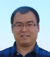
    Dr. Xiaohua Qian is a research fellow at Wake Forest School of Medicine, USA. He obtained Ph.D. from Jilin University, China in 2012. From 2010-2012, Dr. Xiaohua Qian worked as a research scholar in the department of radiology, Duke Medical Center. After that, he was a Research Assistant Professor in Shanghai Advanced Research Institute, Chinese Academy of Science, and then was an Associate Professor in Jiangnan University, China. Dr. Xiaohua Qian has developed many imaging processing, and machine learning algorithms, such as level set segmentation methods, classification-oriented dictionary scheme, and longitudinal multitask sparse regression model. Dr. Xiaohua Qian having extensive experiences in the development of computer-aided diagnosis systems, such as early ischemic stroke detection in CT, cardiac functional analysis, and glioblastoma diagnosis, with the purpose of addressing the clinical challenging. Currently, Dr. Xiaohua Qian working on exploring the radiomics markers and biomarkers for glioblastoma using the radiogenomics and translational studies, integrating the medical imaging, bioinformatics, and clinical research, which bridge the gap between the radiographic phenotype, genotype, and clinical assessments.
  • Ali Gholamrezanezhad, MD, FEBNM

    Department of Diagnostic Radiology
    Case Western Reserve University
    United States
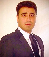
    Dr. Ali Gholamrezanezhad received his Doctorate degree in Medicine (MD) from Tehran University of Medical Sciences, Iran. In 2012 he worked as Postdoctoral Fellowship/Research Scholar at Division of Interventional Radiology, The Russell H. Morgan Department of Radiology and Radiological Science, Johns Hopkins Medical Institutions, Johns Hopkins University, Baltimore, MD, USA and then he completed the Preliminary Internal Medicine Residency program at Yale New Haven Hospital, Yale School of Medicine, New Haven, CT, USA. Currently, Dr. Ali Gholamrezanezhad doing Diagnostic Radiology Residency at University Hospitals of Cleveland, Case Western Reserve University, Cleveland, Ohio, USA. He is a Member of the Scientific Subcommittee for American Roentgen Ray Society (AARS) and American Society of Emergency Radiology (ASER).
  • Yingkun Guo, MD

    Vice Director, Professor, Department of Radiology,
    Sichuan University
    China (中国)
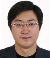
    Prof. Guo is the vice director of Department of Radiology at Sichuan University. His research focuses on cardiovascular imaging, pediatric radiology, molecular and metabolic imaging. As an experienced radiologist, Dr. Guo’s research achievements were published in international professional journals, such as RADIOLOGY, European Radiology, and Journal of Cardiac Magnetic Resonance. Also, Professor Guo is the PI of Molecular Imaging Lab. The laboratory of molecular imaging was established in 2015, and aim to develop novel molecular imaging in vivo, targeted nano-molecular probe, radiogenomics, and imaging contrast mechanisms; evaluate emerging MRI sequences in animal models of human diseases; translate and apply innovative diagnostic imaging in clinic.
  • Yu-Hsiang Juan, MD

    Chang Gung Memorial Hospital and Chang Gung University
    Assistant Professor, Department of Medical Imaging and Intervention
    Taiwan (台灣)
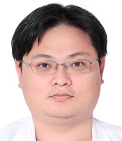
    Dr. Yu-Hsiang Juan is currently an assistant professor and a radiologist in Chang Gung Memorial Hospital, Linkou, Taiwan. He received his medical training from China Medical University and graduated on 2006. He received 4 years of resident training as a radiologist in Chang Gung Memorial Hospital, which he became an attending physician on 2010. Between 2012 to 2014, he was enrolled and completed a 2-years research fellowship training specialized in cardiovascular imaging from the Non-invasive Cardiovascular Imaging Program in Brigham and Women’s Hospital. During the visit in the USA, he also received a Level II Certificate of Cardiovascular CT Experience from Society of Cardiovascular Computed Tomography. He returned to Chang Gung Memorial Hospital in 2014 and awarded the position as an assistant professor since 2015. Currently he has 46 SCI publications, in which he was first, shared first author or corresponding author on 31 of them. He was also a qualified instructor for post-graduate year student and Teaching On The Run (TOTR) Instructor in Chang Gung Memorial Hospital.
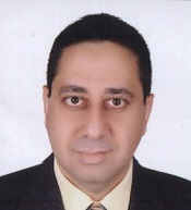
  • Ahmed Abdel Khalek, MD

    Mansoura University
    Professor, Department of Diagnostic Radiology
    Egypt (‫مصر‬‎)
    Prof. Ahmed Abdel Khalek Abdel Razek received his Master degree of Diagnostic Radiology from Mansoura University in 1989 and received MD degree of Diagnostic Radiology from Mansoura University in 1994. Currently he is working as Professor of diagnostic radiology at Mansoura faculty of medicine from July 2004 till date. He is an active member of many associations and also supported as organizing committee member for many international conferences and meetings. He won national award certificate from Mansoura University in 2010. He works as lead author, published 100 scientific papers at peer-reviewed high impact journals at PubMed and 40 scientific papers at national journals. He wrote 4 book chapters and presented more than 350 scientific paper and educational exhibit at international meetings of radiology (RSNA, ECR, ASNR, ASHNR and ARRS). He is reviewer at 35 high impact journals and in the editorial board of 7 international journals and societies. He was supervisor on 80 PhD and MD thesis. He won scientific awards from RSNA and ECR during his career. Research fields are focused on head and neck imaging, neuroradiology and advanced magnetic resonance imaging techniques such as diffusion MR imaging, diffusion tensor imaging, perfusion MR imaging, MR spectroscopy and contrast enhanced MR angiography. In addition, he has some work at computed tomography and color Duplex ultrasound.
  • Polyane Mazucatto Queiroz, MS

    Department of Dental Radiology
    University of Campinas
    Brazil (Brasil)
    Polyane Mazucatto Queiroz, a researcher and dentist, graduated in Dentistry from the State University of Londrina (Londrina, PR - Brazil) (2013) where she developed research and final paper in Dental Radiology, with emphasis on digital radiographic imaging and digital radiographic subtraction technique. Has a Master's Degree in Dental Radiology from the Piracicaba Dental School, State University of Campinas (FOP-UNICAMP) (2015), Piracicaba, SP, Brazil. In the master's degree, worked with Cone Beam Computed Tomography (CBCT) images, studying artifacts in tomographic images, artifact reduction tools and the quality of CBCT images. Currently, she is a doctoral student in Dental Radiology at the same institution where she studied the master's degree. She is working with evaluation of digital radiography sensors (CCD, CMOS and CdTe), evaluating image quality, diagnosis and dosimetry. Also, she has been publishing papers in international journals and developing research involving radiographic images and diagnosis in correlated areas, micro computed tomography (image quality, artifacts and artifact correction tool in microCT), study of the technical aspects of CBCT images and tools of CBCT equipment. She has clinical experience in the area of head and neck diagnostic imaging.

Assistant Editors

    Coming Soon...

Early View

Current Issue

Volume 1, Issue 1

Archive