Optimum operating conditions are an integral part of a successful microvascular surgery. We hereby introduce a new technique to make a lowcost microsuction cannula, which will be helpful for microvascular surgeons.
Video 1. Low-cost microsuction cannula in microvascular surgery. This video demonstrates the assembly of a cost-effective microsuction cannula, essential for maintaining a clear, blood-free surgical field in microvascular procedures. Employing a suction tube, a 2 ml syringe, and a cannula sheath, this approach provides an efficient solution for surgeons in resource-constrained environments, ensuring the successful survival of free flaps through impeccable anastomoses.
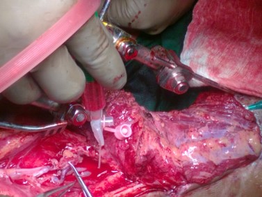
Figure 1. A microsuction cannula used during surgery.

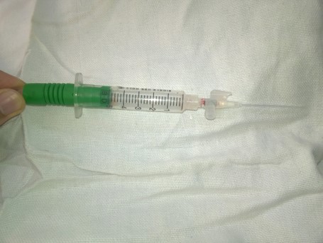
Figure 2. A microsuction cannula made from a syringe and an intravenous cannula.
1. Jacobson JH 2nd, Suarez EL. Microvascular surgery. Dis Chest 1962;41:220-224. PMID: 14450733
2. Acland RD, Sabapathy SR. Acland's practice manual for microvascular surgery. Third Edition 2008 Indian J Plast Surg. 2008;41(2):247. PMCID: PMC2740510
Received date: June 01, 2018
Accepted date: June 17, 2018
Published date: July 08, 2018
None
None
© 2018 The Author(s). This is an open-access article distributed under the terms of the Creative Commons Attribution 4.0 International License (CC-BY).

Video 1. Low-cost microsuction cannula in microvascular surgery. This video demonstrates the assembly of a cost-effective microsuction cannula, essential for maintaining a clear, blood-free surgical field in microvascular procedures. Employing a suction tube, a 2 ml syringe, and a cannula sheath, this approach provides an efficient solution for surgeons in resource-constrained environments, ensuring the successful survival of free flaps through impeccable anastomoses.
This article is interesting and can be published in its current form. The author is suggested to provide with a short video.
ResponseThank you for your suggestion. We have provided with a video.
Kumar P, Ajai KS. A low cost microsuction tip cannula in microvascular surgery. Int Microsurg J 2018;2(1):2. https://doi.org/10.24983/scitemed.imj.2018.00067
 PDF
PDF