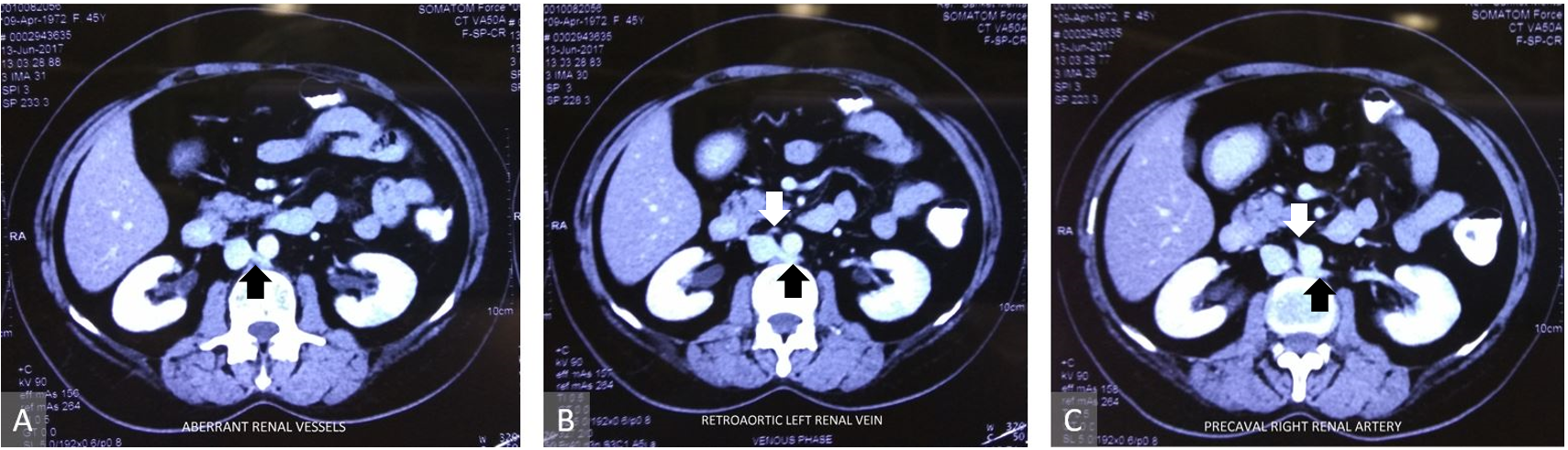
A 55-year-old postmenopausal lady presented with recurrent carcinoma of ovary. A computed tomography scan of the abdomen was performed after neoadjuvant chemotherapy, which showed retroaortic left renal vein and precaval right renal artery (Figure 1), along with the resolution of previous vaginal vault lesion. The intraoperative findings revealed a peritoneal carcinomatosis index of 3, according to an incidental deposit found on the vaginal vault infiltrating into the rectum. During retroperitoneal lymph node dissection, as a part of the procedures in cytoreduction, aberrant renal vessels were identified. A complete clearance was done safeguarding the vessels as seen in the intraoperative image (Figure 2).

Figure 1. Contrast enhanced computed tomography images. Panel A, B, and C show the course of retroaortic left renal vein (black arrows). Precaval right renal artery is shown with the white arrow in Panel B and C.
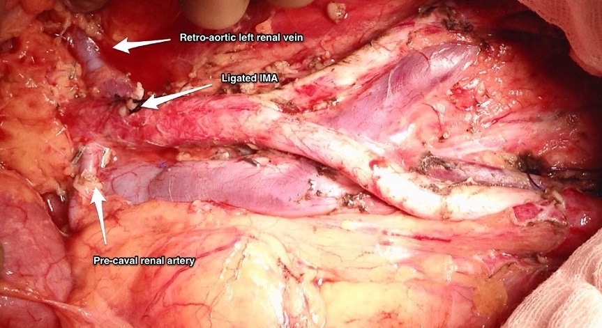
Figure 2. Aberrant renal vessels.
Received date: November 21, 2017
Accepted date: June 24, 2018
Published date: February 20, 2019
None
None
© 2019 The Author (s). This is an open-access article distributed under the terms of the Creative Commons Attribution 4.0 International License (CC-BY).

Waghoo S, Kammar P, Mehta S. Aberrant renal vessels. Ann Case Rep Images 2019;2(1):2. https://doi.org/10.24983/scitemed.acri.2019.00099
 PDF
PDF