Thoracic sympathectomy has been performed for more than a century, primarily for regional hyperhidrosis and selected autonomic or ischemic disorders. Endoscopic thoracic sympathectomy (ETS) can be highly effective for palmar, craniofacial, axillary, and plantar hyperhidrosis. However, some patients develop disabling adverse effects, most notably compensatory sweating below the denervated level, thermoregulatory dysfunction, and excessive dryness above the denervated region. These complications may worsen over time and substantially impair quality of life. Medical therapy is often ineffective for refractory symptoms, and subsequent surgical modifications at different sympathetic levels have produced inconsistent outcomes, prompting interest in reversal strategies. Surgical reversal of ETS aims to restore anatomic continuity of the sympathetic trunk. Early endoscopic reconstruction techniques involved direct coaptation or interposition grafting with vein or nerve autografts, often secured with fibrin glue. Outcomes were unpredictable because of limited surgical access, extensive scarring from previous ETS, and the technical difficulty of intrathoracic microsurgical nerve repair. Robotic platforms have since enabled more precise intrathoracic microsurgery and suture-based nerve coaptation. Since the initial reports of robotic sympathetic trunk reconstruction (STR) in 2016, the surgical team at Chang Gung Memorial Hospital has developed a standardized robotic microsurgical approach with progressively less invasive techniques and refinements in donor-nerve harvesting. Clinical experience suggests that recovery after STR may be prolonged and multiphasic, involving neural reconnection, reinnervation, recruitment or reactivation of sympathetic pathways, and longer-term adaptation of sympathetic function. The following clinical narrative describes the prolonged and potentially systemic course of recovery after robotic STR. I was the 33rd patient to undergo reversal surgery at Chang Gung Memorial Hospital in Taiwan. By 5 years after STR, my compensatory sweating had diminished markedly, and my thermoregulatory function had improved. Because the compensatory sweating that developed after the initial ETS was severe, my primary goal in pursuing reversal surgery was relief of these symptoms. During recovery, however, I experienced several unexpected systemic changes. These observations reflect a single first-person experience rather than formal outcome data. They may offer insight into the complexity of the sympathetic nervous system, the broader physiologic effects associated with sympathectomy, and the gradual adaptation that may occur after restoration of sympathetic trunk continuity.
Evolution of Sympathectomy for Hyperhidrosis
Sympathetic surgery dates to the late 19th century. Alexander reported the first sympathectomy in 1889 for the treatment of epilepsy, and in 1920, Kotzareff reported the first sympathectomy for hyperhidrosis [1]. Primary hyperhidrosis, particularly palmar hyperhidrosis, is now the principal indication for endoscopic thoracic sympathectomy, although selected patients with axillary or craniofacial hyperhidrosis may also undergo the procedure [1,2]. Beyond hyperhidrosis, thoracic sympathetic ablation has occasionally been used in carefully selected patients with facial blushing, angina, arrhythmias, complex regional pain syndrome (CRPS), and certain upper-limb ischemic disorders, including Raynaud phenomenon [3].
Endoscopic technique and complications
The standard contemporary approach is endoscopic thoracic sympathectomy (ETS). After identification of the sympathetic trunk and ganglia, sympathetic interruption is achieved by resection, clipping, or electrocoagulation, typically between the second and fourth rib levels. This approach can provide effective relief of palmar hyperhidrosis and other regional forms, including craniofacial, axillary, and plantar hyperhidrosis. However, some patients develop excessive postoperative anhidrosis above the targeted level. Compensatory sweating may also develop below the denervated region. These changes can disrupt thermoregulation and may persist or become difficult to manage over time. In affected patients, these complications can substantially impair quality of life [4].
Rationale for surgical reversal
Nonoperative measures, including pharmacologic therapy, are often ineffective for severe postoperative symptoms. Secondary surgical interventions have therefore been explored, including division of the lower thoracic sympathetic trunk and subsequent lumbar sympathectomy. However, reported outcomes have been inconsistent and remain controversial. Surgical reversal procedures have consequently been introduced in an attempt to restore sympathetic continuity [5,6].
Emergence of ETS Reversal Surgery
Surgical reversal of ETS, through sympathetic trunk reconstruction (STR), was initially proposed to mitigate the adverse effects of sympathectomy by restoring anatomic continuity of the sympathetic trunk. A major operative challenge is the deep intrathoracic location of the sympathetic trunk, which limits surgical exposure and access. Published experience with STR remains limited. Historically, sympathetic trunk defects were reconstructed endoscopically by direct coaptation or by interposition grafting with vein, intercostal nerve, or sural nerve grafts, typically secured with fibrin glue [5].
Limitations of Endoscopic Approaches
Although the endoscopic approach avoids the morbidity associated with thoracotomy, clinical outcomes have been variable. Conventional endoscopic instruments provide limited precision for microsurgical suturing, leading to reliance on fibrin glue for graft fixation. Without suture-based coaptation, maintaining precise alignment and stability of the neural repair may be challenging. In addition, identification and preparation of viable nerve stumps require microsurgical expertise and may be difficult in the presence of scarring from previous sympathectomy. These technical limitations may contribute to inconsistent outcomes. Consequently, purely endoscopic STR has been increasingly replaced by approaches that permit more precise microsurgical reconstruction [5–7].
Robotic Microsurgery for ETS Reversal
In 2016, Connery reported the first three cases of robotic STR, two of which had favorable outcomes [8]. In 2017, a multidisciplinary team at Chang Gung Memorial Hospital in Taiwan, comprising specialists in reconstructive microsurgery and robotic thoracic surgery, established a robotic STR program. The first patient was a 60-year-old woman who had undergone bilateral T2–T4 sympathectomy 25 years earlier. She presented with compensatory sweating below the nipple line, thermoregulatory dysfunction, and anhidrosis of the head, neck, and both upper extremities. She also reported fatigue, avolition, and depressive symptoms.
Initial clinical implementation
The patient underwent robotic STR using a sural nerve autograft. Nerve coaptation was performed with microsurgical sutures rather than fibrin glue, providing direct fixation and anatomic continuity of the reconstructed nerve. At 1 year, the patient reported substantial improvement in her symptoms. Following this initial experience, robotic STR for ETS reversal was introduced routinely at the institution in January 2019.
Program expansion and refinement
As of February 2026, the institutional cohort included 185 patients, 66 of whom were international referrals. One patient underwent revision STR with the current technique after a previous reversal procedure had failed. The surgical protocol has continued to evolve. The current standardized technique uses a vascularized intercostal nerve as the donor graft, and adoption of a single-port robotic system has further reduced the invasiveness of the procedure. Early cases in the series, including my own, required bilateral thoracic incisions and sural nerve harvesting, whereas the current procedure requires only two incisions [9–15]. Robotic STR has subsequently been adopted by surgeons in the United States, China, France, and India, with some approaches incorporating techniques developed by the Chang Gung group.
At the time of reversal surgery on September 29, 2020, I was a 57-year-old man who had undergone bilateral thoracic sympathectomy in 1993, 27 years earlier, for severe bilateral palmar hyperhidrosis. According to the 1993 operative report, the sympathectomy extended from T2 through T5 on the left and from T2 through T6 on the right.
Although I am a practicing radiologist, I did not fully understand the complexity of the sympathetic nervous system at the time of either the original sympathectomy or the later reversal. My understanding of the changes that occur after sympathectomy and reversal is still evolving, but I believe that my experience may be useful to both patients and clinicians in understanding the prolonged and often multistage course of recovery.
Prereversal Sweating Pattern
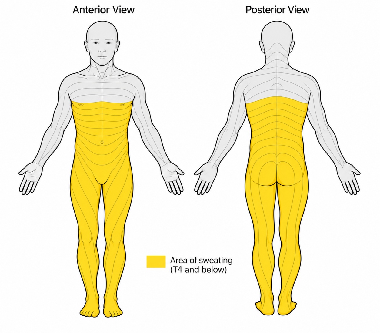
Before reversal surgery in September 2020, I had a distinct regional pattern of sweating (Figure 1). The area above the nipple line was persistently anhidrotic and remained completely dry, whereas the areas below the nipple line showed severe compensatory sweating that had gradually worsened over the years after sympathectomy.
Functional Impact and Surgical Decision
The compensatory sweating substantially affected my daily life and limited my ability to continue marathon running. I had previously qualified for the Boston Marathon five times. As my thermoregulatory dysfunction and compensatory sweating progressed, I began exploring surgical options. In 2020, I learned of the early clinical experience with reversal surgery at Chang Gung Memorial Hospital. I contacted the hospital and discussed the procedure with the surgical team. I then decided to undergo reversal surgery.
Preoperative Evaluation
The operation was performed during the coronavirus disease 2019 (COVID-19) pandemic. I arrived in Taiwan 22 days before surgery and underwent testing for severe acute respiratory syndrome coronavirus 2 (SARS-CoV-2). After a negative test result, my wife and I completed a 15-day hotel quarantine, followed by 7 days in a hotel near the hospital. I then met with the surgical team, including T.N.J. Chang and Y.K. Chao. The operative report from my original 1993 ETS was reviewed for surgical planning.
To establish a preoperative baseline, I rated the severity of my symptoms using the Chang Gung Sympathetic Function Questionnaire (CG-SFQ) [4]. Individual symptoms were rated on a 0–10 numeric rating scale, with 0 indicating no symptoms and higher scores indicating greater symptom severity. A medical history was also obtained, including smoking history, systemic disease, and previous surgical procedures.
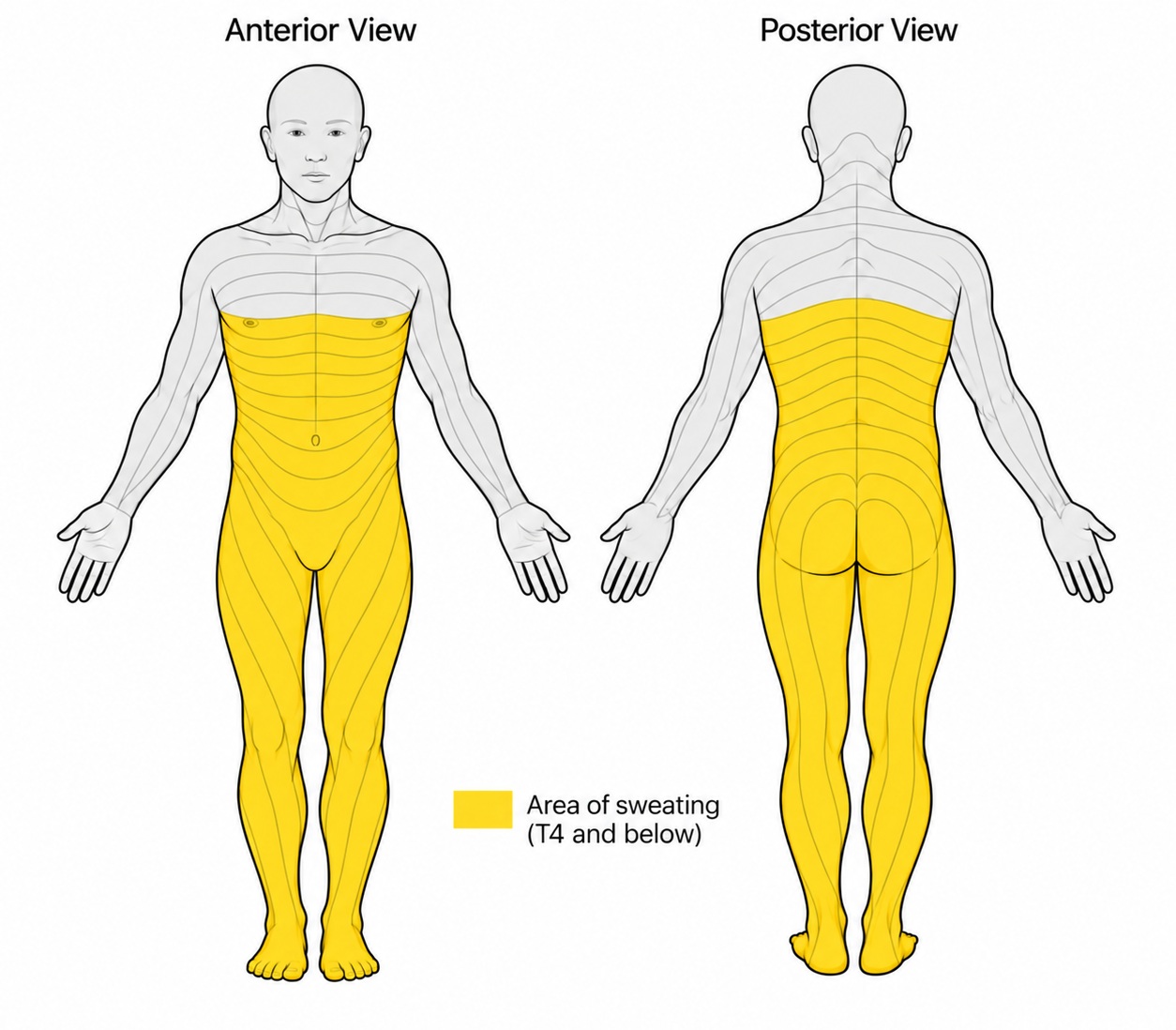
Figure 1. Prereversal distribution of compensatory sweating and dermatomal reference in September 2020. Anterior and posterior body diagrams show the reported sweating pattern before sympathetic trunk reconstruction. Yellow shading denotes areas of compensatory sweating extending from approximately the T4 level caudally over the trunk and lower extremities, whereas the region above this level remained anhidrotic and dry. Superimposed dermatomal contours are provided as an anatomic reference to illustrate the spatial relationship between the reported sweating distribution and segmental cutaneous innervation.
I was the 33rd patient to undergo this procedure at Chang Gung Memorial Hospital. I was admitted the day before surgery and taken to the operating room the following morning. Standard patient identification procedures were completed before surgery.
Anesthesia and Patient Positioning
After induction of general anesthesia and intubation with a double-lumen endotracheal tube, I was placed in the semiprone position with the right side down and appropriate padding and protection. The left chest wall and left lower extremity were prepared and draped. A pneumothorax was established with carbon dioxide insufflation at a pressure of 6 to 8 mm Hg.
Thoracic Port Placement
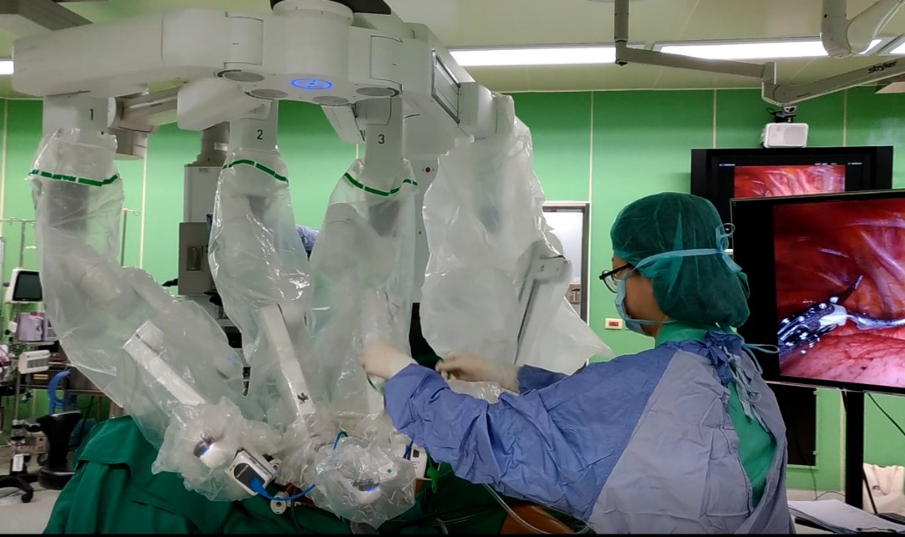
Thoracic ports were then placed (Figure 2). A 30° thoracoscope was inserted into the seventh intercostal space along the posterior axillary line. The right robotic arm was positioned in the fifth intercostal space along the posterior axillary line, medial to the scapula. The left robotic arm was placed in the ninth intercostal space along the posterior axillary line. An assistant port was placed in either the sixth or seventh intercostal space along the midaxillary line.
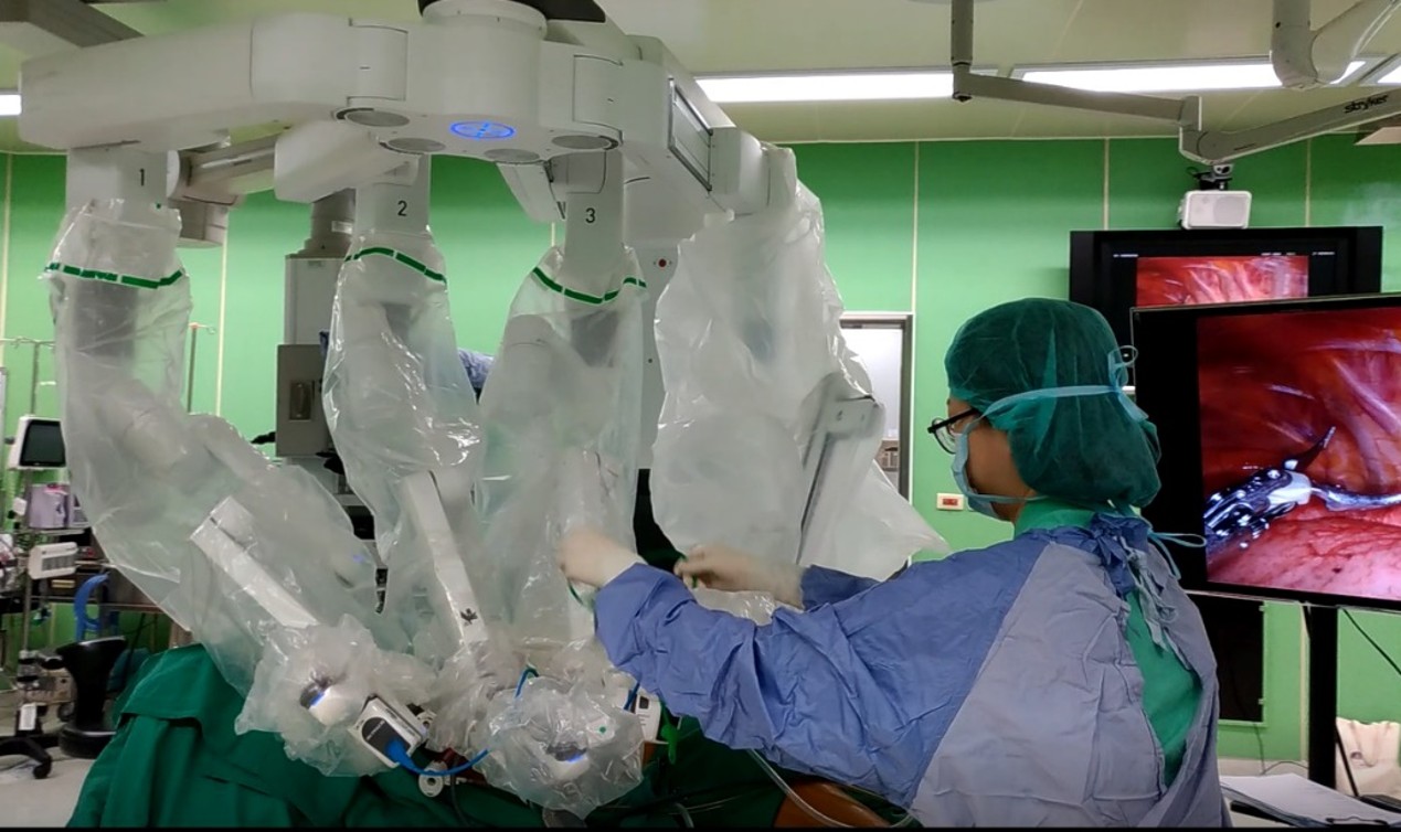
Figure 2. Robotic setup for left-sided sympathetic trunk reconstruction. The patient was positioned semiprone with the right side down, and the da Vinci Xi robotic system was docked to provide left-sided thoracic access. The robotic arms were positioned over the operative field while the surgical team initiated the thoracic phase of the procedure. The intrathoracic operative view was displayed on the adjacent monitor, permitting visualization of the surgical field during robotic dissection and subsequent reconstruction.
Robotic System Docking
The da Vinci Xi surgical system (Intuitive Surgical, Inc., Sunnyvale, CA, USA) was docked at a 45° counterclockwise angle relative to the patient’s craniocaudal axis. Once docked, the robotic platform provided magnified three-dimensional visualization and articulated instrument movement for the subsequent intrathoracic dissection and microsurgical preparation of the sympathetic trunk.
Sympathetic Trunk and Graft Preparation
A two-team approach was used. The thoracic surgery team explored the thoracic cavity and prepared the sympathetic trunk for reconstruction, while the reconstructive microsurgery team simultaneously harvested the left sural nerve from the ankle to the knee. The harvested sural nerve was prepared as the autologous graft for subsequent STR.
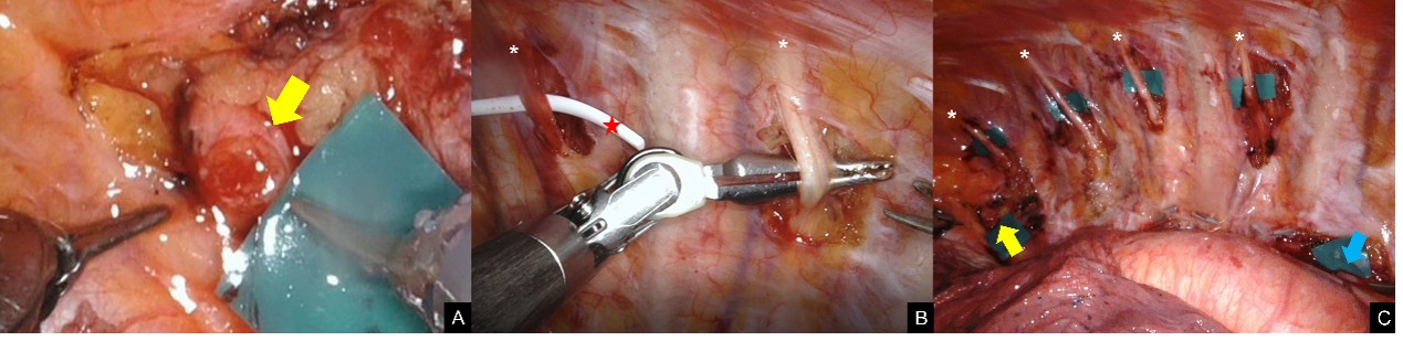
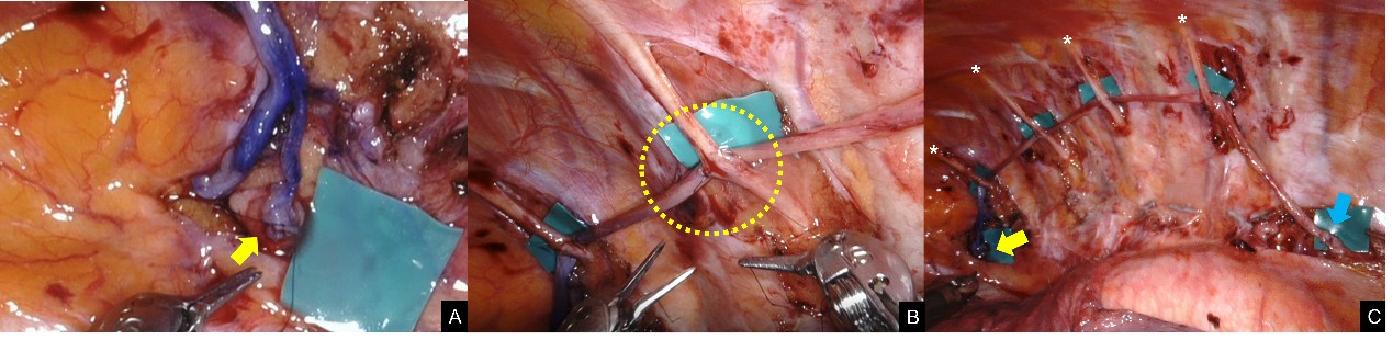
After entering the thoracic cavity, the thoracic surgery team lysed existing adhesions and identified the proximal and distal sympathetic trunk stumps and adjacent intercostal nerves (Figure 3). Scarring from the previous ETS was dissected with Potts scissors, monopolar scissors, and a curved bipolar dissector. Scar tissue surrounding the sympathetic trunk stumps was carefully released to avoid excessive dissection or injury to the nerves.
The proximal stump was generally larger than the distal stump because the sympathetic trunk progressively decreases in caliber below the stellate ganglion. The sympathetic trunk was exposed to the inferior margin of the stellate ganglion and dissected until pliable proximal and distal nerve stumps were identified. Stump viability and fascicular integrity were assessed visually. Fresh bleeding and a mushrooming appearance at the cut surface were considered intraoperative indicators of stump viability (Figure 3A). Care was taken to avoid injury to the adjacent subclavian vessels and aorta.
After viable proximal and distal nerve stumps had been prepared, the required graft length was measured. The graft was selected to be 20% longer than the measured defect to allow tension-free coaptation.
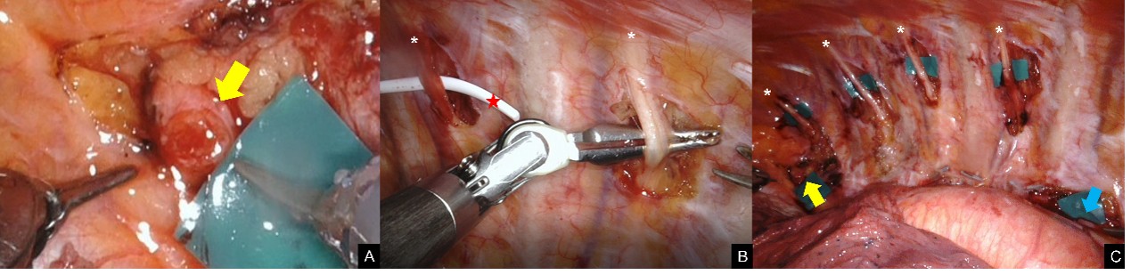
Figure 3. Preparation of the left sympathetic trunk and adjacent intercostal nerves for reconstruction. (A) Following microsurgical dissection, the proximal sympathetic trunk stump, corresponding to the distal portion of the stellate ganglion (yellow arrow), appeared soft and pliable, with mushrooming and fresh bleeding at the cut surface, findings considered intraoperative indicators of stump viability. (B) A white vessel loop (red star) was passed beneath the involved left intercostal nerves (ICNs; asterisks) to facilitate their identification and preparation for reconstruction. (C) The prepared proximal sympathetic trunk stump (yellow arrow), distal stump (blue arrow), and adjacent left ICNs from T2 through T5 (asterisks) are shown before nerve graft coaptation.
Sural Nerve Harvest
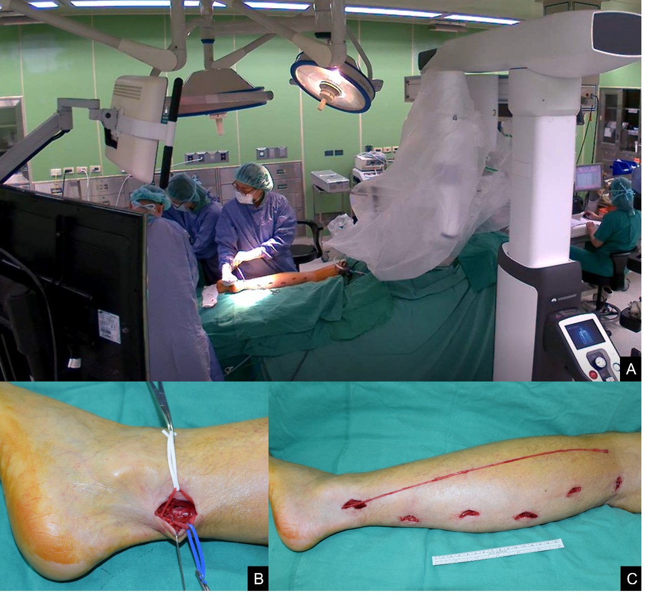
While the thoracic surgery team performed adhesiolysis and sympathetic trunk dissection, the microsurgery team simultaneously harvested the sural nerve (Figure 4A). The sural nerve was harvested from the left leg because this was expected to interfere less with driving during recovery. A stepwise multiple-incision technique was used. The initial 3- to 5-cm incision was made posterior to the lateral malleolus at the retrofibular level to identify the sural nerve (Figure 4B). The sural nerve lies deep to the lesser saphenous vein and immediately posterior to the posterior border of the fibula. Harvest proceeded from distal to proximal along the course of the nerve through sequential incisions, with the most proximal incision made in the popliteal crease (Figure 4C).
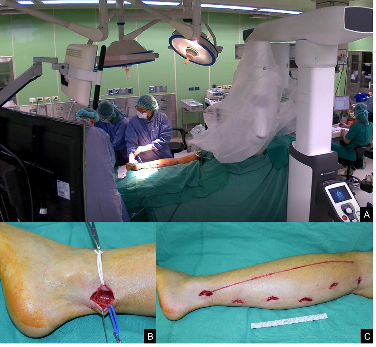
Figure 4. Sural nerve harvest during robotic sympathetic trunk reconstruction. (A) A coordinated two-team operative approach was used. The microsurgery team harvested the left sural nerve from the lower extremity while the thoracic surgery team simultaneously performed left-sided thoracic adhesiolysis and sympathetic trunk dissection using the robotic platform. This parallel workflow allowed donor-nerve preparation to proceed concurrently with preparation of the thoracic reconstruction field. (B) The sural nerve was identified through an initial 3- to 5-cm retrofibular incision placed posterior to the lateral malleolus. The lesser saphenous vein is encircled with a blue vessel loop, whereas the deeper sural nerve is encircled with a white vessel loop, allowing clear differentiation of the adjacent structures before proximal dissection. (C) Harvest then proceeded from distal to proximal through a series of stepwise incisions along the course of the sural nerve. The harvested sural nerve is shown laid along the posterolateral lower leg, with a ruler provided for scale. The sequential incisions permitted progressive mobilization of the nerve, with the most proximal exposure extending to the popliteal crease before proximal transection and preparation of the autologous graft for subsequent sympathetic trunk reconstruction.
Graft length determination
At the popliteal crease, the posterior tibial and common peroneal nerves were palpated for anatomic verification before proximal transection of the sural nerve. To achieve tension-free coaptation within the thorax, the required graft length was calculated as 1.2 times the distance between the stellate ganglion and the inferior margin of the sympathectomy defect. In an adult, 35 to 40 cm of sural nerve can typically be harvested from one leg, and communicating branches may also be harvested if additional length is required. The total graft length used in this case was 28 cm, comprising 13 cm for the left hemithorax and 15 cm for the right hemithorax.
Graft trimming and donor closure
After harvest, the distal end of the sural nerve was marked, and redundant soft tissue surrounding the epineurium was removed on the back table. The leg incisions were closed with polyglycolic acid sutures and reinforced with 2-octyl cyanoacrylate topical skin adhesive (Dermabond; Ethicon, Inc., Raritan, NJ, USA). Dry gauze and an elastic compression wrap were applied to reduce the risk of hematoma or seroma.
Microsurgical Nerve Grafting
Control of the robotic system was then transferred from the thoracic surgery team to the microsurgery team. Particular care was required during reconstruction because of the proximity of the aorta to the robotic instruments.
Left-sided positioning and proximal coaptation
The left-sided reconstruction was performed first with the patient in the semiprone position and the left side up. This position allowed simultaneous sural nerve harvest and thoracic dissection. The sural nerve autograft was used to bridge the proximal and distal sympathetic trunk stumps. Coaptation was performed with microforceps and 8-0 nylon sutures.
Graft routing and distal coaptation
After proximal coaptation was completed (Figure 5A), the graft was routed deep to the involved intercostal nerves toward the distal stump. The graft was cut to the required length, and the remaining segment was preserved for reconstruction of the right side. Distal coaptation was performed in the same manner. Fewer sutures were required distally because of the smaller caliber of the distal sympathetic trunk.
Side-to-side intercostal nerve coaptation
The graft was then secured to each adjacent intercostal nerve with two to three sutures using side-to-side coaptation (Figure 5B). This technique is hypothesized to establish communication between the reconstructed sympathetic trunk and the intercostal nerves, simulating the function of the white and gray rami communicantes. This routing also avoids placement of the graft within the scarred bed of the previous ETS site and helps prevent graft displacement. All coaptations were confirmed to be tension-free.
Hemostasis and chest tube placement
After completion of the left-sided STR (Figure 5C), hemostasis was achieved. A chest tube was inserted through the assistant port under direct endoscopic visualization, with care taken to avoid contact with the neural reconstruction sites. The chest tube was placed to water seal for passive drainage rather than active suction to reduce the risk of mechanical disruption of the nerve coaptations.
Repositioning and right-sided reconstruction
After lung re-expansion, the thoracic incisions were closed with topical skin adhesive. I was then repositioned into the semiprone position with the right side up. The remaining sural nerve graft was introduced into the right thoracic cavity, and the reconstruction was performed in a similar manner. The right-sided procedure was technically less complex because the aorta was not within the operative field.
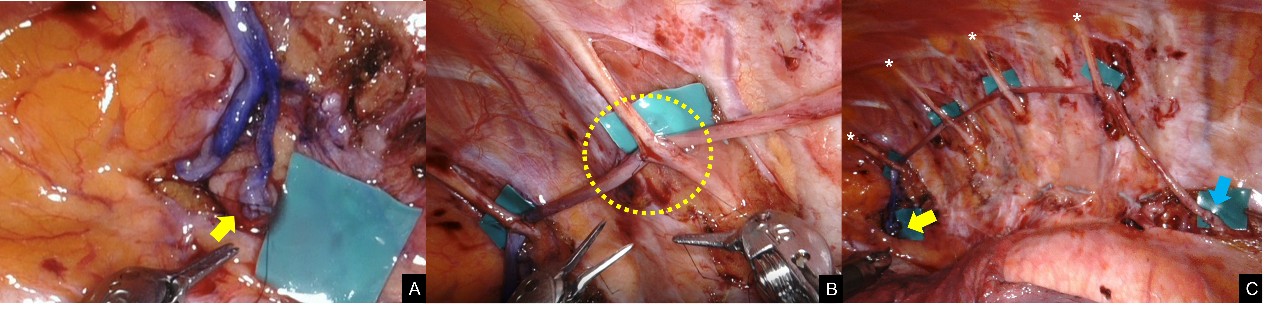
Figure 5. Left-sided sympathetic trunk reconstruction with a sural nerve graft. (A) The ink-marked end of the harvested sural nerve graft was positioned at the proximal left sympathetic trunk stump (yellow arrow) and secured with 8-0 nylon microsutures to establish proximal coaptation. (B) After proximal fixation, the graft was routed along the thoracic wall and coapted side-to-side to the adjacent second through fifth intercostal nerves (ICNs). The yellow circle highlights a side-to-side coaptation site between the sural nerve graft and an adjacent ICN. These sequential coaptations were performed while maintaining the graft along the planned reconstruction course. (C) Final appearance of the completed left-sided reconstruction, with the sural nerve graft spanning the proximal sympathetic trunk stump (yellow arrow) and distal stump (blue arrow). The involved ICNs are indicated by asterisks, demonstrating the relationship of the graft to the adjacent intercostal nerves after completion of the reconstruction.
Postoperative Care
I was extubated in the operating room and transferred to the intensive care unit for 24 hours of respiratory monitoring. Immediate extubation may not be appropriate for all patients, particularly those with a history of smoking or chronic obstructive pulmonary disease [16].
The chest tubes were removed on postoperative day 1, and subsequent chest radiography confirmed the absence of pneumothorax or hemothorax. I was instructed to perform deep-breathing exercises and undergo chest physiotherapy. Ambulation was restricted during the first postoperative week. I was transferred to the general surgical ward on postoperative day 2 and discharged on postoperative day 5.
The compression dressing over the left donor leg was maintained at discharge to reduce the risk of hematoma or seroma. Suture removal and formal physical therapy were not required. I experienced transient chest wall paresthesia, numbness of the plantar and lateral aspects of the left foot, and mild left lower extremity edema, which persisted for several months.
After reversal, I was asked to complete the CG-SFQ at 6-month intervals for up to 5 years after surgery, with available assessments extending through 60 months, as arranged by Dr. Lisa Wen-Yu Chen [4].
Throughout my postoperative course, I remained engaged with patient and medical communities and periodically shared my experience. In October 2021, approximately 1 year after reversal, I participated in a webinar hosted by Chang Gung Memorial Hospital and described my recovery to clinicians and other patients. In February 2025, I participated in an International Microsurgery Club webinar, where I discussed a presentation by T.N.J. Chang and described my longer-term course. In October 2025, at 5 years after reversal, I shared a written account of my postoperative course with an online patient community. These interactions provided opportunities to share my first-person experience of the prolonged and evolving course after reversal with patients and clinicians.
In my experience, some patients appear to expect recovery to occur relatively quickly, and some early postoperative accounts describe the surgery as unsuccessful. I believe that such conclusions may be premature when only a short period has elapsed since surgery. My own prolonged recovery prompted me to examine the possible neurophysiologic processes involved and to share my experience in the hope that it may help patients and clinicians better understand the time course of recovery.
Dermatomal Sweating Evolution
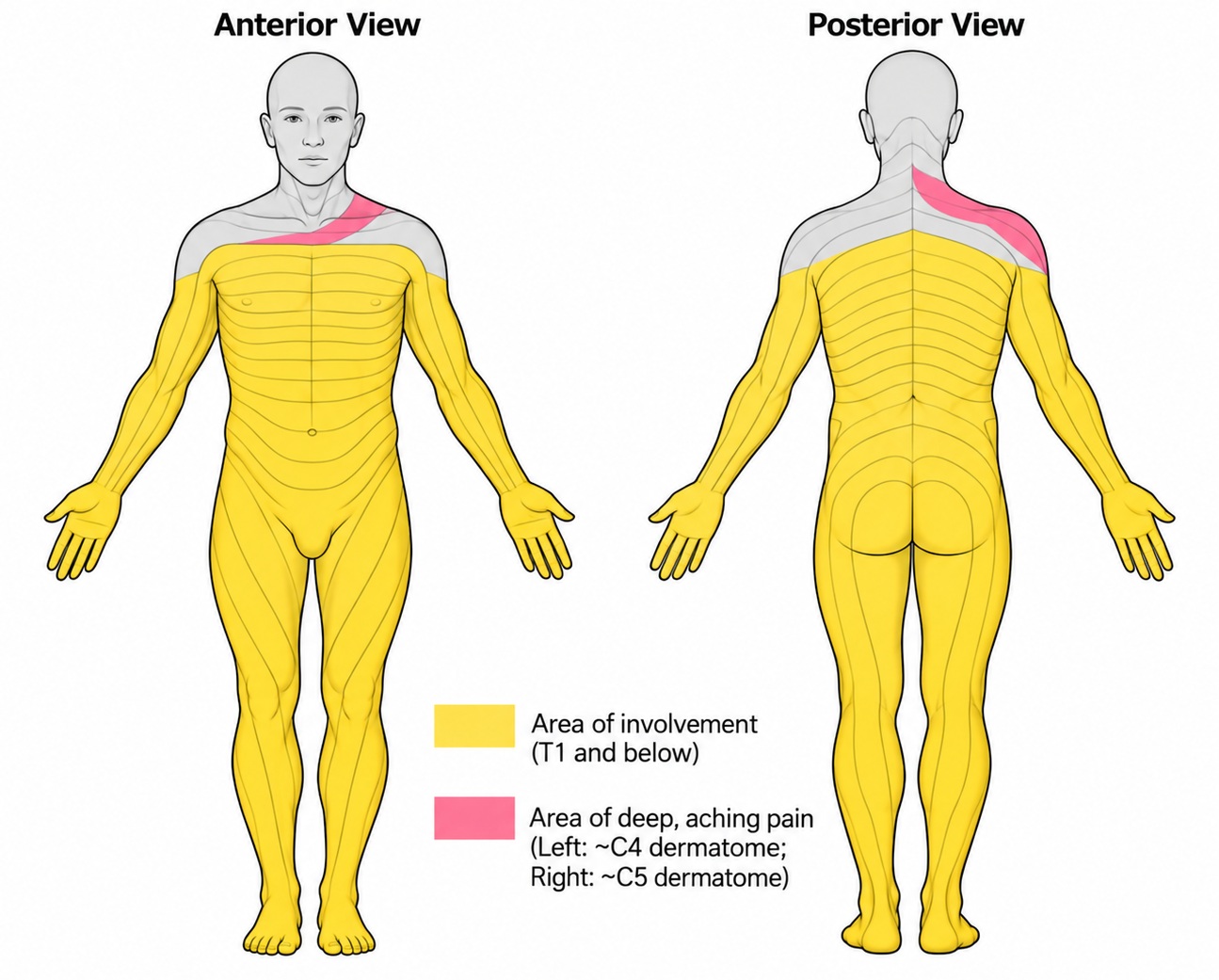
In the months and years after reversal, I noticed a recurring pattern in which a deep, aching pain resembling a muscle cramp developed within a dermatomal distribution approximately 1 to 2 months before sweating appeared at that level. Yellow shading indicates areas of sweating in Figures 1 and 6; in Figure 6, pink shading indicates the location of this pain at 5 years after reversal.
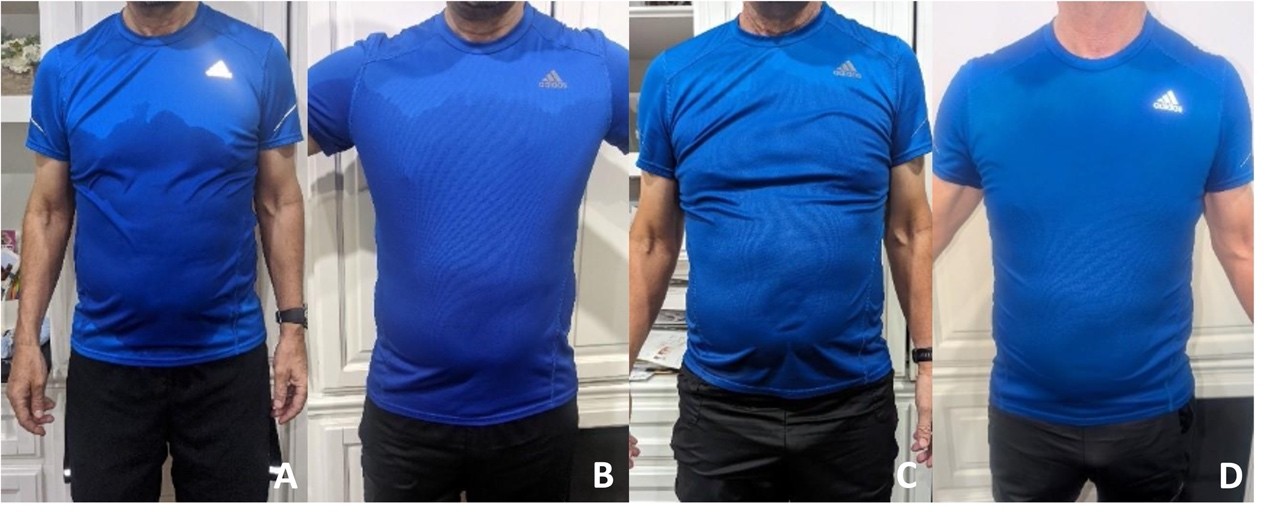
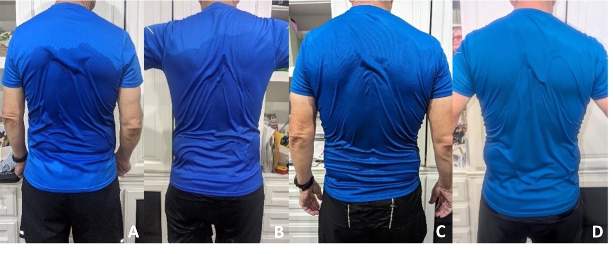
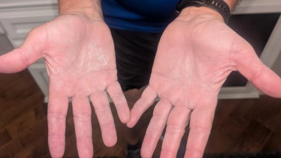
The photographs provide only a partial representation of the change because the overall amount of sweating during exercise has also decreased substantially. Before reversal, after 30 minutes on an elliptical machine followed by 30 minutes on a stationary bicycle, I often had to wipe a large pool of sweat from the floor. I now have little or no sweat dripping onto the floor during comparable strenuous exercise, and some areas of sweating no longer soak through my shirt. Postexercise photographs showing the evolution of my anterior and posterior sweating patterns from the prereversal baseline through 1, 3, and 5 years after reversal are shown in Figures 7 and 8. The return of palmar sweating at 5 years is shown in Figure 9.
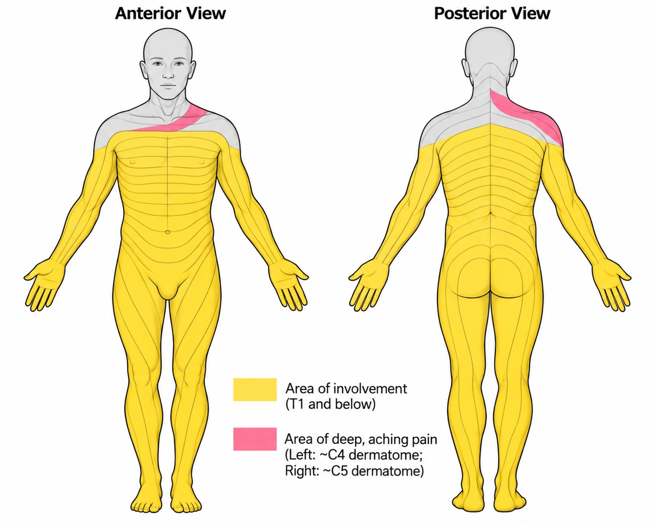
Figure 6. Distribution of sweating and deep aching pain 5 years after sympathetic trunk reconstruction. Anterior and posterior schematics depict the reported distribution of sweating and localized deep aching pain in September 2025, approximately 5 years after reversal surgery. The yellow-shaded region, labeled in the figure as “Area of involvement (T1 and below),” denotes the reported distribution of sweating at that time; sweating in the more cephalad regions remained relatively mild. Pink shading indicates the areas of deep, aching pain, localized approximately to the C4 dermatome on the left and the C5 dermatome on the right. The pain involved the upper anterior chest and shoulder region anteriorly and extended over the corresponding upper posterior shoulder and parascapular regions. Superimposed dermatomal contours are provided as an anatomic reference to illustrate the spatial relationship between the reported sweating distribution, areas of pain, and segmental cutaneous innervation. The schematic represents the author’s reported symptom distribution at this single postoperative time point and does not constitute direct physiologic mapping of sympathetic reinnervation.
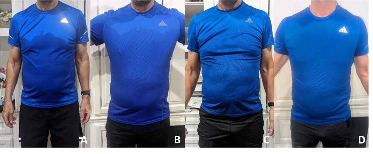
Figure 7. Serial anterior sweat distribution before and after sympathetic trunk reconstruction during exercise. Anterior photographs were obtained after 30 minutes of elliptical exercise followed by 30 minutes on a stationary bicycle to document changes in sweat distribution over time. (A) Before reversal, shirt wetness was concentrated over the lower anterior chest and extended superiorly to just below the nipple line, with relative dryness of the upper chest. (B) At 1 year after reversal, anterior sweating remained predominantly below the upper chest, and the superior margin of visible shirt wetness remained below the shirt logo. (C) At 3 years, a similar distribution persisted, although the pattern of anterior wetness continued to evolve, with the upper boundary remaining below the logo. (D) At 5 years, the superior extent of visible sweating had progressed to the level of the shirt logo, with additional bilateral wetness near the junction of the upper chest and neck. Despite this more cephalad distribution, the overall area and intensity of shirt wetness were less extensive than before reversal.
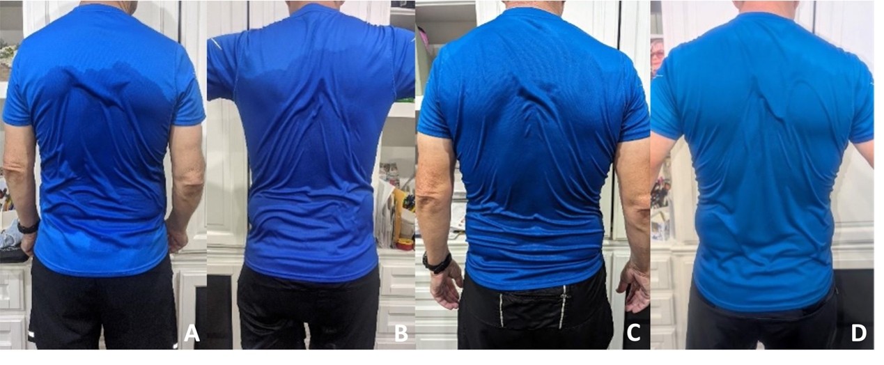
Figure 8. Serial posterior sweat distribution before and after sympathetic trunk reconstruction during exercise. Posterior photographs were obtained after 30 minutes of elliptical exercise followed by 30 minutes on a stationary bicycle to document longitudinal changes in sweating. (A) Before reversal, visible shirt wetness was concentrated predominantly over the mid-to-lower back, with relative sparing of the upper back and shoulder regions. (B) At 1 year after reversal, posterior sweating remained centered over the lower and mid back, with limited superior extension. (C) At 3 years, the upper boundary of visible wetness had progressed cephalad and was higher on the left than on the right. (D) At 5 years, this asymmetric cephalad progression was more apparent, with wetness extending nearly to the shirt collar along the midline and over the left upper back, while the right upper back remained relatively less involved. The serial photographs demonstrate a gradual upward extension of the posterior sweating distribution over the 5-year postoperative period.
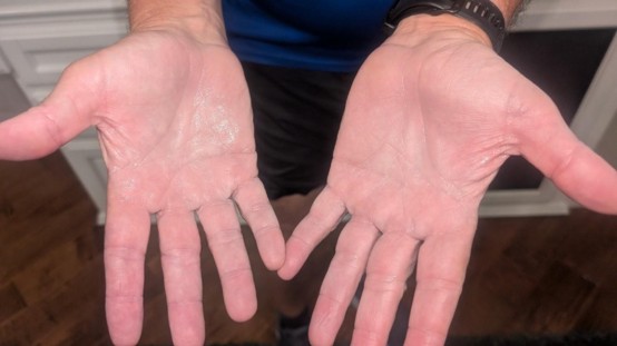
Figure 9. Return of palmar sweating 5 years after sympathetic trunk reconstruction. Photograph obtained after 30 minutes of elliptical exercise followed by 30 minutes on a stationary bicycle demonstrates visible moisture over the palmar surfaces of both hands. Before reversal, palmar sweating had remained absent after the original thoracic sympathectomy, with the previously denervated upper-body region remaining persistently dry. At 5 years after reversal, the presence of bilateral palmar moisture documents the return of sweating in an area that had remained anhidrotic before sympathetic trunk reconstruction.
Thermoregulatory Improvement
The return of sweating in my arms, axillae, upper chest, and upper back has coincided with a substantial improvement in my ability to tolerate heat and thermoregulate. I am much more functional than I was before reversal. However, recovery took considerably longer than I initially expected, and I experienced several unexpected postoperative effects. My experience suggests that recovery after reversal may be prolonged and may involve several stages rather than a simple return of function after nerve grafting.
Initial Sympathectomy and Adaptation
I developed severe palmar hyperhidrosis during medical school and residency. After discussing ETS with a thoracic surgeon, I elected to undergo the procedure in the summer of 1993. At that time, neither of us anticipated the severity of the compensatory hyperhidrosis that would later develop. To address the palmar symptoms, an extensive sympathectomy was performed from T2 through T5 on the left and from T2 through T6 on the right.
For several years after surgery, my palmar sweating remained absent and I was generally able to function normally. I had increased sweating of the lower body and an unusual distribution of sweating soon after surgery, but I could still thermoregulate adequately. Compensatory hyperhidrosis then increased slowly over subsequent years.
Progressive Long-Term Deterioration
My thermoregulatory function became increasingly impaired approximately 10 years after sympathectomy. Over the following 17 years, I developed progressively severe compensatory hyperhidrosis, impaired thermoregulation, and recurrent nocturnal sweating that often limited my sleep to fewer than 4 or 5 hours per night. These symptoms increasingly affected my social, occupational, and personal activities.
The sweating initially involved my chest and back and progressively extended to my legs and buttocks. Eventually, I could not walk my dog for 1 mile at temperatures above 18°C (65°F) without my lower body becoming soaked. At work, even while sitting and reviewing radiologic studies with residents, my back and buttocks could become saturated with sweat. Once sweating began, it often required 30 to 60 minutes to stop. I also became unable to continue many of my usual sporting activities, including tennis, golf, and marathon running. By 27 years after sympathectomy, these symptoms had become severe enough that I was actively seeking a surgical solution.
Recovery Timeline After Reversal
I entered reversal surgery expecting that within approximately 1 year I might recover something close to my pre-1993 sweating pattern. At my postoperative visit 1 week after surgery, Dr. Chang told me that he expected recovery to be more complete at approximately 2 years.
My subsequent course was considerably longer. The most substantial improvement occurred between 4 and 5 years after surgery. This experience led me to view recovery as a prolonged process rather than one determined solely by the time required for axonal regrowth. I hope that this observation may help patients avoid losing confidence during the early stages of recovery and may help surgeons provide more realistic expectations regarding the postoperative course.
Expected Nerve Regrowth
Using the commonly cited clinical estimate of approximately 1 mm/day for peripheral axonal regeneration [12], I estimated that axonal growth across the 15-cm right-sided graft might require approximately 150 days, or 5 months, whereas growth across the 13-cm left-sided graft might require approximately 130 days, or 4.25 months. I therefore initially expected that meaningful function might begin to return within approximately 5 to 6 months. This calculation represents a heuristic estimate rather than a patient-specific measurement of axonal regeneration.
Over time, however, I came to regard this estimate as describing only one component of recovery. To organize my own experience, I divided the postoperative course into four proposed phases.
Phase 1: neural reconnection
The first phase was the immediate surgical reconstruction performed on September 29, 2020, when the sural nerve grafts were used to restore anatomic continuity between the proximal and distal sympathetic trunk stumps.
Phase 2: axonal reinnervation
The second phase represents axonal growth through the sural nerve grafts. Using the same approximate clinical estimate of 1 mm/day [12], I calculated that axonal growth across the 15-cm right-sided graft and 13-cm left-sided graft might require approximately 150 and 130 days, respectively. I therefore expected axonal growth to reach the distal stumps within approximately 4 to 5 months after surgery.
Phase 3: reactivation and recruitment
I use the third phase to describe the period beginning approximately 6 months after surgery, when I began to experience changes that I interpreted as reflecting reactivation and recruitment of sympathetic pathways. During this period, my compensatory sweating and thermoregulation gradually improved, but several unexpected symptoms also developed.
These included skin pain, discoloration, skin thickening, hair loss, new varicose veins of the chest wall and legs, and lower extremity swelling with pitting edema. I also experienced changes involving my right eye in the setting of preexisting amblyopia and longstanding central serous chorioretinopathy. Many of these findings have subsequently improved or nearly resolved.
Phase 4: autonomic recalibration
I use the term recalibration to describe the prolonged phase during which clinical changes continued to evolve as sympathetic function appeared to recover. This is a conceptual framework based on my clinical course rather than a directly measured neurophysiologic process.
In my case, the painful and inflamed areas of skin that developed during the earlier phase began to improve substantially only after approximately 4 years. The lower extremity edema has resolved, and the hair loss involving my axillae and scalp vertex has almost completely recovered.
In areas of residual rash and discoloration of the upper body, I continue to notice a deep, cramping pain within a dermatomal distribution before sweating develops at that level. At approximately 5 years after reversal, this pain was centered at C4 on the left and C5 on the right (Figure 6). As sweating has appeared progressively at more cephalad levels, my thermoregulation has also continued to improve. Episodes of spontaneous sweating involving the buttocks, back, and abdomen under ordinary conditions have become substantially less frequent. These changes have occurred over months and years rather than over the days or weeks I initially expected.
Postoperative Expectations
Patients considering reversal should be aware that restoration of anatomic continuity does not necessarily correspond to immediate functional recovery. In my case, the surgical reconstruction was completed successfully, but the subsequent clinical changes evolved over several years.
I initially assumed that axonal growth through the graft would largely determine the recovery period. The unexpected symptoms that began approximately 6 months after surgery led me to consider a broader and more complex process. The four-phase framework described here represents my interpretation of a single postoperative course and should not be regarded as an established model of sympathetic nerve recovery.
Systemic Cutaneous Changes
My first major diffuse cutaneous reaction began approximately 6 months after surgery. I abruptly developed a diffuse maculopapular rash associated with darkened, thickened skin and intense pruritus. Initially, I suspected an allergic reaction or, less likely, a fungal infection. The rash and itching extended from my scalp to my toes.
I was evaluated by family medicine and dermatology physicians and was treated with antifungal and topical glucocorticoid therapy, without improvement. Over the following month, the acute pruritic eruption evolved into diffuse skin thickening and hyperpigmentation accompanied by varicose veins of the chest wall and legs and hair loss involving the axillae, legs, and scalp vertex.
The affected skin subsequently became painful, with burning and pins-and-needles sensations resembling the discomfort of sunburn. These changes were not confined to the previously denervated upper body but extended to my legs and feet. I also developed swelling of the hands and lower extremities, particularly from the calves to the toes. The abnormalities persisted for several years but began to improve approximately 4 years after surgery, particularly around the axillae and flanks.
CRPS-Related Mechanistic Considerations
These findings led me to consider whether some features of my postoperative syndrome resembled reflex sympathetic dystrophy, now termed CRPS. CRPS typically develops after a localized injury and is associated with abnormalities of pain processing, vasomotor control, sudomotor function, inflammation, and regional autonomic regulation [17,18].
A number of mediators have been implicated in CRPS, including catecholamines, cytokines, substance P, bradykinin, and nitric oxide [18,19]. These pathways can influence pain, vascular tone, edema, inflammation, and cutaneous function [18,19]. C fibers are also involved in nociception, temperature sensation, itch, sudomotor responses, and autonomic regulation of the skin [20].
Although some of my postoperative findings resembled features described in CRPS, my diffuse presentation does not establish that the same pathophysiologic process was present.
Nerve-Stem Cell Crosstalk
Another potentially relevant concept is crosstalk between nerves and stem cells in skin disease and cutaneous nerve regeneration [21]. Interactions among cutaneous nerves, hair follicles, arrector pili muscles, stem cells, neuropeptides, vasculature, and immune pathways may influence skin and hair homeostasis.
Such interactions provide one possible framework for considering the combination of pain, pigmentary change, altered vascularity, edema, and hair loss that I experienced. Whether these mechanisms contributed directly to my postoperative findings remains unknown.
Proposed C-Fiber Mechanism
I initially interpreted my cutaneous symptoms as evidence that previously altered neural pathways were becoming active again after reversal. I considered whether sprouting and new synaptic activity involving afferent and efferent C fibers might contribute to the simultaneous appearance of pain, sweating, vascular changes, and altered skin sensation.
In this framework, the symptoms could reflect imperfect coordination between newly active peripheral pathways and central autonomic regulation. As sympathetic function returned, some areas developed more appropriate sweating and improved thermoregulation, whereas others were associated with pain, rash, edema, or vascular changes.
This interpretation remains speculative because the underlying neural activity was not measured directly.
Gradual Cutaneous Improvement
As recovery progressed, the rash and skin pain gradually improved. Some areas near my axillae and anterior chest returned to normal-appearing skin, whereas other areas continued to change more slowly. I interpreted this gradual improvement as consistent with ongoing adaptation of the neural and autonomic responses involved in sweating, pain, vascular tone, and sensation. The precise mechanism remains uncertain. These changes occurred gradually over several years as sweating patterns and thermoregulatory function continued to evolve.
Improvement in Compensatory Sweating
My compensatory hyperhidrosis has improved gradually each year since reversal, although much more slowly than I initially expected. I now sweat in my axillae, upper back, thorax, arms, and hands.
Soon after reversal, I continued to have severe lower-body sweating while playing golf in our desert climate. My shorts and lower shirt would become completely soaked, and sweat would run down my legs and form a pool beneath me at the driving range. I now golf several times each week and rarely have sweat pouring down my legs, even at temperatures above 32°C (90°F). I still sweat heavily below the nipple line in hot conditions, but the sweating now stops much more quickly when I enter an air-conditioned room or vehicle.
Postexercise photographs also demonstrate progressive upward extension of sweating compared with my prereversal and early postoperative pattern. During 30 minutes of exercise on an elliptical machine followed by 30 minutes on a stationary bicycle, I previously produced enough sweat to form a large pool beneath the equipment and completely soak a towel. With comparable exercise now, little or no sweat reaches the floor.
A similar change has occurred with running. Before reversal, my shoes became completely saturated during runs of 5 to 10 miles and often required 1 to 2 days to dry. My wife bought a shoe-drying machine because the shoes frequently remained wet between training sessions. They are now only mildly damp after a run.
Functional Thermoregulatory Recovery
For me, the location of sweating alone is not the most important measure of recovery. My primary goals were to reduce inappropriate sweating at ordinary ambient temperatures and to regain the ability to stop sweating promptly after heat exposure or exercise.
Both have improved. I can now work with residents without routinely soaking the back of my shirt or the chair on which I am sitting. I can walk my dog at approximately 27°C (80°F) without needing to change all of my clothing afterward. After exercise, sweating now generally stops within approximately 5 minutes rather than continuing for as long as an hour. These improvements developed gradually over several years.
Five-Year Cutaneous Recovery
At 5 years after nerve reconstruction, the cutaneous abnormalities have also begun to resolve more clearly. Improvement is most apparent around the T4 and T5 dermatomes on the left and T5 and T6 on the right. Skin color has returned toward normal in these regions, and the pain and varicose veins are improving near the axillae and anterior chest. I expect that additional changes may continue beyond 5 years, although the ultimate duration and extent of recovery remain uncertain.
Ocular Clinical Course
The sympathetic nervous system contributes to the regulation of multiple organ systems, and my postoperative experience also included ocular changes. I was born with amblyopia of the right eye without strabismus. The amblyopia was not detected until approximately 6 years of age and resulted primarily in reduced depth perception.
Approximately 6 years after sympathectomy, I developed diplopia while playing sports, particularly tennis. An ophthalmologist could identify no clear cause and recommended that I stop correcting the amblyopic eye. I subsequently wore a contact lens only in the stronger left eye, which reduced the diplopia.
Approximately 10 years after sympathectomy, I developed loss of central vision in the right eye and was given a diagnosis of central serous chorioretinopathy on fluorescein angiography. Central serous chorioretinopathy is characterized by serous subretinal fluid accumulation associated with retinal pigment epithelial dysfunction and hyperpermeability of the underlying choroid [22]. At the time, my ophthalmologist noted that the condition is often associated clinically with stress-related factors, although I did not identify such a trigger in my case.
Postreversal Ocular Course
One week after reversal, I developed right-sided Horner syndrome. An obvious right-sided lid lag persisted for approximately 3 years before gradually improving, with only minimal residual asymmetry at present.
Approximately 1 year after surgery, I also began to experience recurrent episodes of severe dizziness, right eye pain, and nausea. These episodes occurred several times per week and sometimes required me to lie down or remain motionless with my eyes closed until the symptoms resolved. They occasionally affected me while driving or interpreting radiologic studies.
Approximately 3 years after reversal, I began to notice improvement in central vision in the right eye after nearly 20 years of a central visual field defect. At approximately the same time, the dizziness and lid lag began to improve. I have now had no episodes of dizziness for approximately 18 months. Improvement in central visual acuity was subsequently confirmed by an optometrist, and I resumed wearing a contact lens in the right eye with some improvement in depth perception and peripheral vision.
Ocular Autonomic Interpretation
I have considered whether the ocular changes that occurred before and after reversal might be related in some way to autonomic regulation in an eye already affected by amblyopia. I also considered whether the postoperative Horner syndrome, episodes of dizziness, and later improvement in central vision might have accompanied broader autonomic changes during recovery.
These temporal associations do not establish a causal relationship between sympathectomy, reversal surgery, amblyopia, central serous chorioretinopathy, or subsequent visual recovery. My ocular course may be unique because of the preexisting amblyopia and central serous chorioretinopathy. Several ocular findings evolved during the same period as my postoperative recovery, although their relationship to sympathetic recovery remains uncertain. These observations reinforced my impression that the clinical changes after sympathectomy and reversal may extend beyond sweating alone, but their underlying mechanisms remain unknown.
Longitudinal Questionnaire Findings
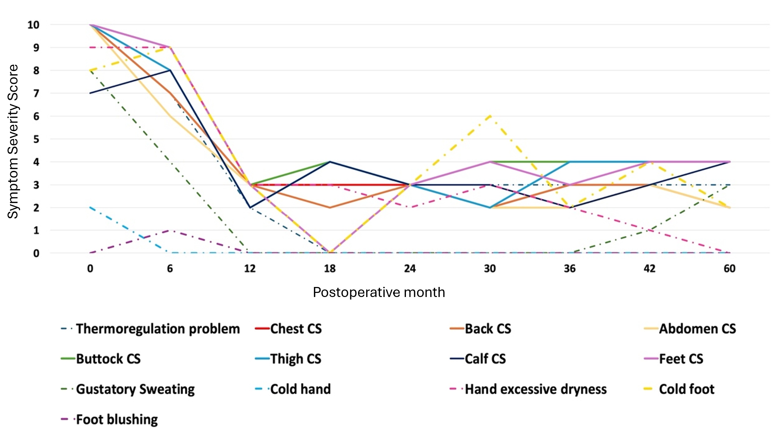
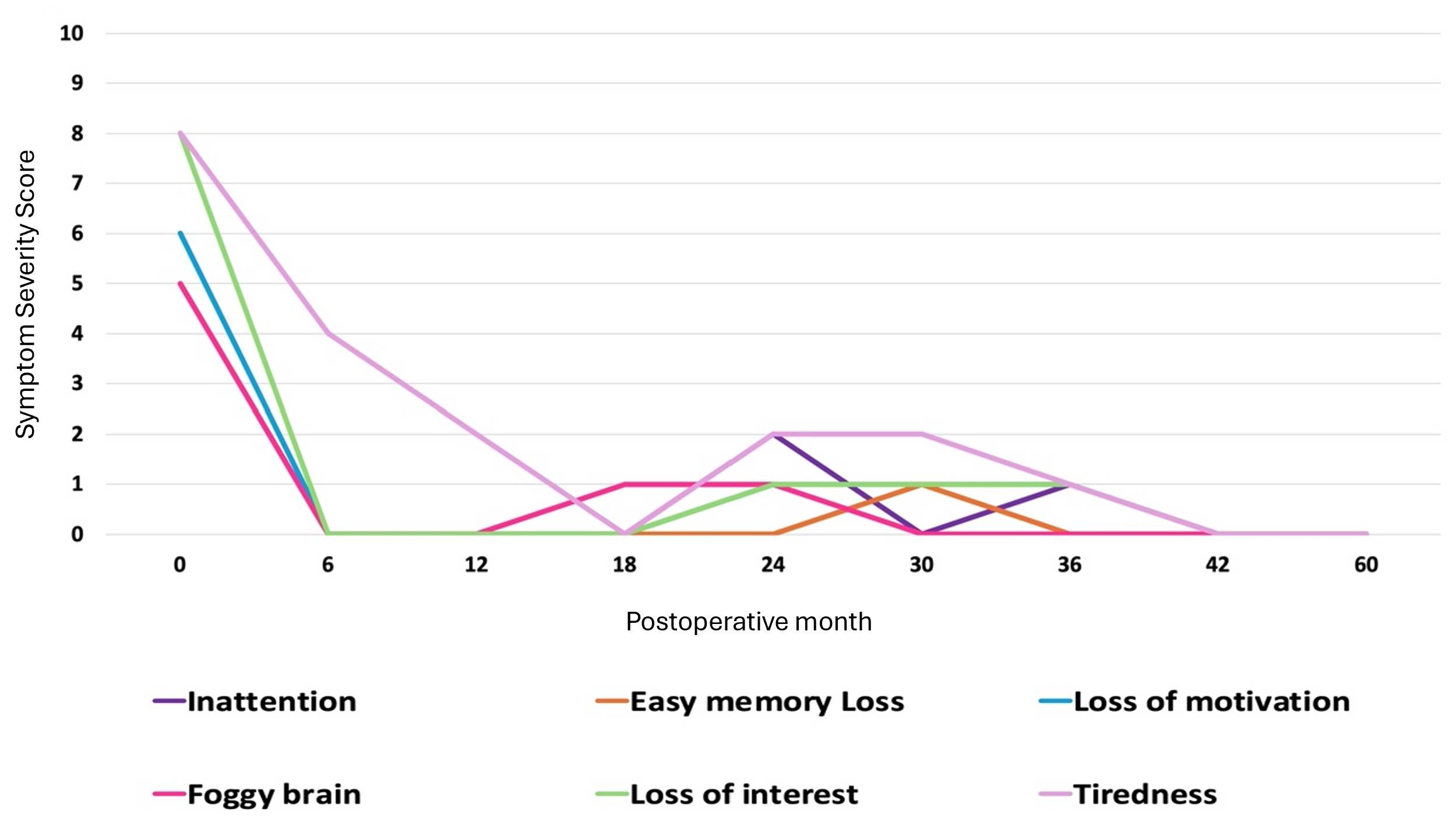
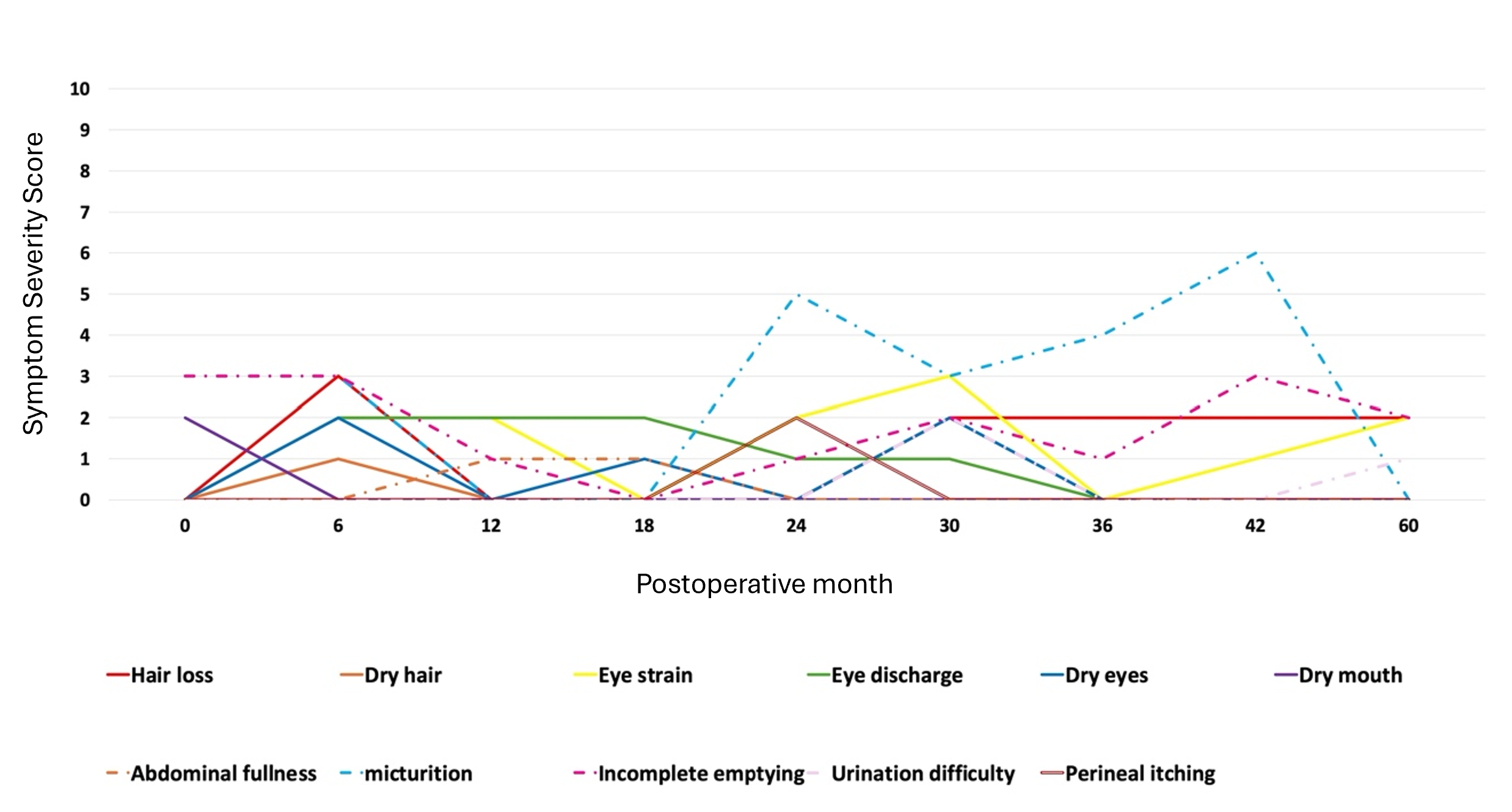
Serial CG-SFQ scores provided a structured longitudinal record of self-reported symptoms during the 5-year postoperative period (Figures 10–12). Figure 10 summarizes compensatory sweating, thermoregulatory symptoms, and other autonomic symptoms. Figure 11 summarizes self-reported cognitive and related symptoms, whereas Figure 12 summarizes selected somatic symptoms. Figures 11 and 12 provide complementary information on additional cognitive and somatic symptom domains, contributing to a more complete longitudinal record of the postoperative course. These scores represent serial self-reported ratings from a single individual and were not accompanied by objective physiologic measurements or formal statistical analysis.
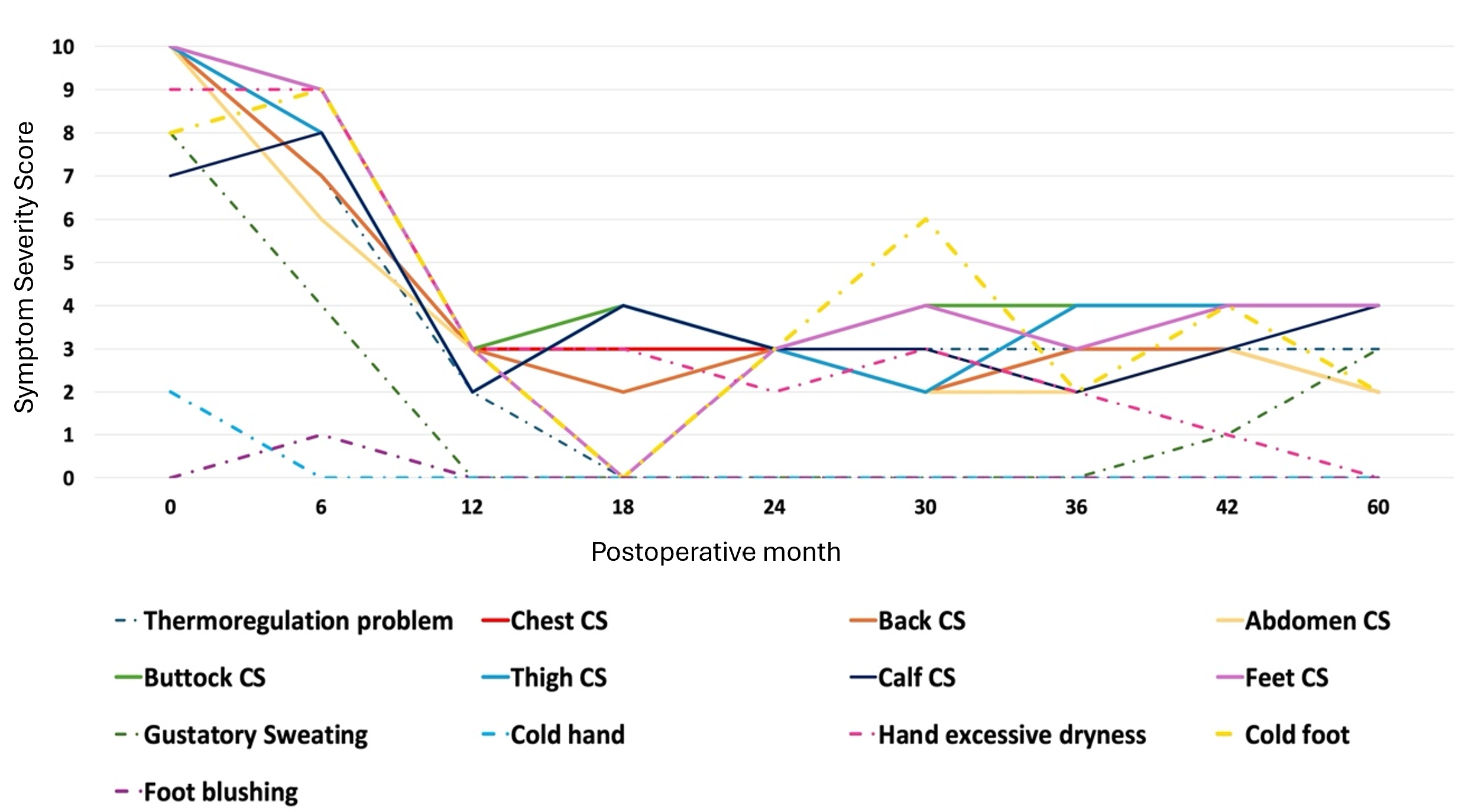
Figure 10. Longitudinal self-reported symptom severity scores before reversal and during postoperative follow-up. Symptoms were assessed using the Chang Gung Sympathetic Function Questionnaire (CG-SFQ) on a 0–10 numeric rating scale, with 0 indicating no symptoms and higher scores indicating greater symptom severity. Baseline scores were obtained before reversal, with available postoperative assessments extending through 60 months. Month 0 represents the prereversal baseline. Serial scores are shown for thermoregulatory problems; compensatory sweating (CS) involving the chest, back, abdomen, buttocks, thighs, calves, and feet; gustatory sweating; cold hands; excessive hand dryness; cold feet; and foot blushing. CS, compensatory sweating.
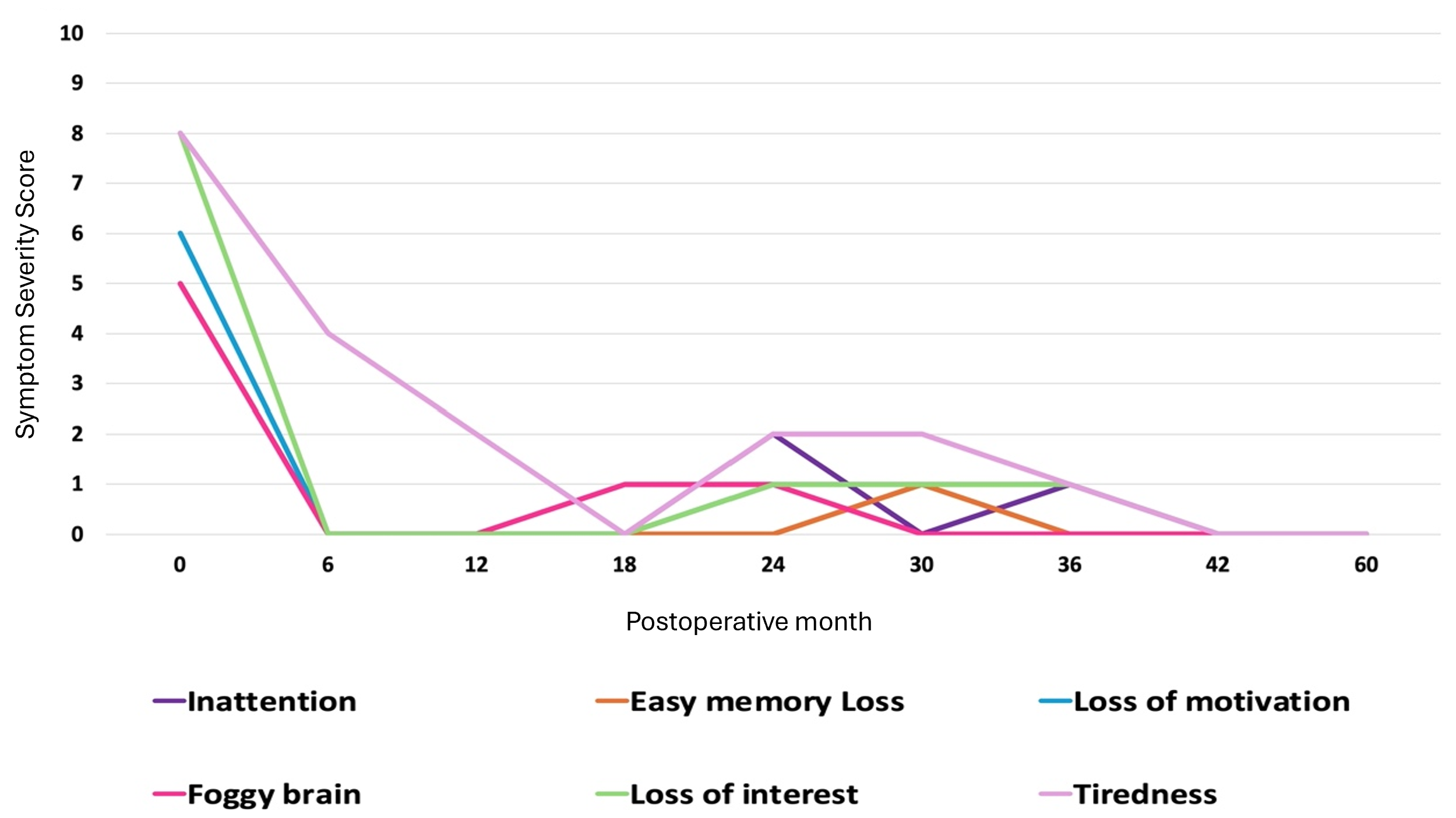
Figure 11. Longitudinal self-reported cognitive and related symptom severity scores before reversal and during postoperative follow-up. Symptoms were assessed using the Chang Gung Sympathetic Function Questionnaire (CG-SFQ) on a 0–10 numeric rating scale, with 0 indicating no symptoms and higher scores indicating greater symptom severity. Baseline scores were obtained before reversal, with available postoperative assessments extending through 60 months. Month 0 represents the prereversal baseline. Serial scores are shown for inattention, easy memory loss, loss of motivation, foggy brain, loss of interest, and tiredness.
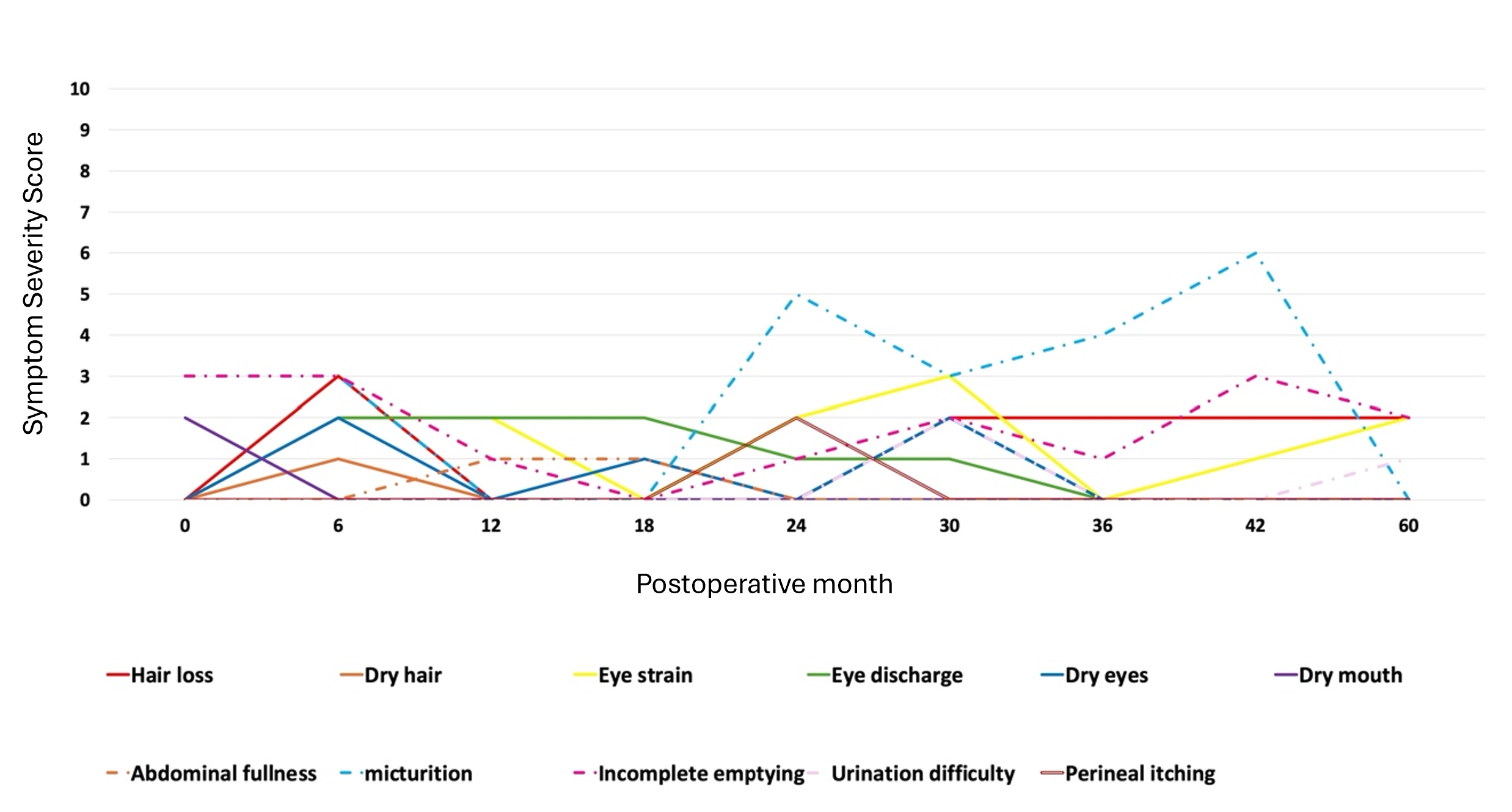
Figure 12. Longitudinal self-reported selected somatic symptom severity scores before reversal and during postoperative follow-up. Symptoms were assessed using the Chang Gung Sympathetic Function Questionnaire (CG-SFQ) on a 0–10 numeric rating scale, with 0 indicating no symptoms and higher scores indicating greater symptom severity. Baseline scores were obtained before reversal, with available postoperative assessments extending through 60 months. Month 0 represents the prereversal baseline. Serial scores are shown for hair loss, dry hair, eye strain, eye discharge, dry eyes, dry mouth, abdominal fullness, micturition, incomplete emptying, urination difficulty, and perineal itching.
This case describes prolonged recovery after robotic STR performed 27 years after extensive bilateral endoscopic thoracic sympathectomy for palmar hyperhidrosis. Severe compensatory sweating and thermoregulatory dysfunction had progressively impaired daily activities and exercise tolerance. Robotic STR with sural nerve autografts restored anatomic continuity of the sympathetic trunks using microsurgical suture-based coaptation. Postoperatively, sweating gradually returned to previously anhidrotic regions, including the palms, while compensatory sweating and heat intolerance improved over several years, with the greatest functional improvement occurring between 4 and 5 years. Published experience with STR remains limited, and the prolonged course observed here extends the usual emphasis on axonal regrowth alone. This case suggests that restoration of anatomic continuity may precede functional recovery by years and highlights the importance of long-term follow-up when evaluating outcomes after STR.
Long-Term Recovery
My course after the initial sympathectomy also evolved slowly. Although the initial sympathectomy involved multiple thoracic levels, I was not severely impaired until approximately 10 years after surgery. Thereafter, my symptoms worsened progressively over the following 17 years, and by 27 years after sympathectomy, immediately before reversal, my functional impairment had become severe.
The prolonged deterioration after sympathectomy raises the possibility that recovery after reversal may likewise occur gradually over an extended period. In my case, the greatest improvement occurred several years after reconstruction, and I continue to experience incremental improvement.
Clinical Implications of ETS
In my clinical radiology practice at a university teaching hospital in the United States, I continue to encounter patients with a history of sympathectomy. The clinical consequences of ETS may extend beyond altered sweating alone, although the nature and mechanisms of these changes remain incompletely defined. Greater recognition of the complexity of sympathetic function and the possibility of prolonged postoperative changes may improve preoperative counseling and shared decision-making. Long-term observations after reversal may also help characterize the trajectory of functional recovery after reconstruction. Robotic sympathetic trunk reconstruction can restore anatomic continuity of the sympathetic trunk, but the relationship between anatomic repair and subsequent functional recovery remains uncertain and may evolve over several years.
Generalizability and Future Directions
Not all patients undergoing sympathectomy reversal are likely to follow the same postoperative course. The CRPS-like features I experienced may not occur in other patients, and my ocular findings may have been influenced by preexisting amblyopia and central serous chorioretinopathy. Accordingly, these observations should be interpreted as a single longitudinal clinical experience rather than as evidence of a uniform pattern of recovery.
Future studies should examine whether demographic and clinical factors, including age, sex, the interval between sympathectomy and reversal, and sympathetic trunk defect length, are associated with the rate or extent of recovery. Larger longitudinal studies using standardized outcome measures could help clarify these relationships and improve patient selection, postoperative counseling, and expectations regarding the duration and extent of recovery.
This single first-person account describes 5-year recovery after robotic STR performed 27 years after extensive ETS. Anatomic continuity was restored, but functional recovery was gradual. Compensatory sweating and heat tolerance improved over several years, with the greatest functional change occurring between 4 and 5 years. This course extended well beyond the period expected from axonal growth across the grafts alone. Cutaneous, vasomotor, and ocular findings also evolved during the same period, although their relationship to sympathetic recovery remains uncertain. These observations do not establish a uniform recovery pattern or a measured mechanism. The proposed phases of reconnection, reinnervation, recruitment or reactivation, and recalibration remain an interpretive framework. Anatomic repair should therefore not be equated with immediate functional recovery. Longer follow-up and comparative studies are needed to determine whether sympathetic trunk defect length and the interval between sympathectomy and reversal are associated with the rate or extent of recovery. Such data may also improve counseling of patients considering reconstruction.
I thank the medical team at Chang Gung Memorial Hospital, Linkou Medical Center, Taiwan, particularly Drs. Yin-Kai Chao, Tommy Nai-Jen Chang, and Lisa Wen-Yu Chen, for their valuable input. I also thank the Chang Gung team for providing the surgical photographs and questionnaire graphs summarizing CG-SFQ symptom scores.
Received date: April 20, 2026
Accepted date: May 12, 2026
Published date: October 30, 2026
Portions of the author’s postoperative experience were previously discussed in educational webinars and shared with an online patient community. The present manuscript has not been previously published or presented in its current form.
This article presents a first-person account of the author’s own clinical experience and does not report the enrollment of additional human participants. The manuscript was prepared in accordance with applicable ethical principles, including those reflected in the Declaration of Helsinki, with appropriate attention to privacy and confidentiality. As the author is also the subject of this report, separate consent to participate was not applicable.
This work received no specific grant from any funding agency in the public, commercial, or not-for-profit sectors. The author conducted the work using personal resources and received no external financial support. The absence of external funding did not influence the design, conduct, interpretation, or reporting of this work.
In accordance with the ethical standards of the SciTeMed Publishing Group, the author declares that there are no financial or other conflicts of interest that could influence the content or interpretation of this work. The author further confirms that no other individual or entity meets the criteria for authorship or has made a substantial contribution warranting authorship.
It is imperative to acknowledge that the opinions and statements articulated in this article are the exclusive responsibility of the author and do not necessarily reflect the views or opinions of the author’s affiliated institutions, the publisher, editors, or reviewers. Furthermore, the publisher does not endorse or guarantee the accuracy of any statements made by the manufacturers or the author. This disclaimer emphasizes the importance of respecting the author’s autonomy and ability to express his own opinions regarding the subject matter, while readers should exercise their own judgment in interpreting the information provided. The author’s position and level of expertise in the subject area should be considered, together with critical evaluation of the information presented. Accordingly, readers should approach the information in this article with an open and discerning perspective.
© 2026 The Author. The article presented here is openly accessible under the terms of the Creative Commons Attribution 4.0 International License (CC BY 4.0). This license permits the use, distribution, and reproduction of the material in any medium, provided that appropriate credit is given to the original author, the copyright holder, and the journal of publication, and that the original source is properly cited. Any use, distribution, or reproduction of the material must comply with the terms of the CC BY 4.0 license. We encourage the use and dissemination of this material in a manner that respects the intellectual property rights of the original author and copyright holder and adheres to accepted standards of citation and attribution in academic publishing.
The reviewer comments and author responses are presented below. Editorial notes have been added to highlight relevant content in the final published article and to supplement the author’s responses where helpful. These notes are based on the final published article and are distinct from the author’s responses.
After consideration of the reviewers’ reports and the manuscript as submitted, I find that the paper addresses a clinically interesting topic but is not yet ready for publication in its current form. Its main strength lies in the long-term postoperative perspective provided by a physician who is also the patient, which draws attention to the prolonged and complex course of recovery after robot-assisted sympathetic trunk reconstruction (STR). However, substantial concerns remain regarding article classification, causal interpretation, neurophysiological precision, data-source separation, and factual accuracy. These issues currently limit the manuscript’s scientific reliability and overall suitability for publication. I am therefore recommending revision and resubmission. A revised version may be reconsidered if the author is able to address the reviewers’ major concerns and improve the manuscript’s clarity, discipline, and evidentiary restraint.
Author responseThe document I sent you was initially a post I sent to the Chang-Gung Linkou reversal Facebook group at my 5-year post-op mark. I was motivated to share my experience by a desire to share with other patients earlier than me in their postoperative recovery and those considering reversal what my understanding of the complexity of the reversal process was and what possible side effects could be encountered after reversal. There is quite a bit of misinformation in the chat group, and people can have unrealistic expectations of a possible quick fix for their ETS side effects. Others post and tell patients that there is no hope for improvement and our situation is not fixable. I shared my post with Tommy Chang. He told me that he felt this was something that would be publishable in the medical literature, which was never my intent. I sent him my document, and he inserted some of the information from their experience that they had previously published. This resulted in a mishmash of a case report and a review article. I have tried to excise the review components and limit it to my case. I am a teaching radiologist at an academic institution, but I do not publish medical articles, so my experience in this space is limited. I do not pretend to fully understand the exact mechanism of my unexpected skin manifestations, but I do know it is a systemic process that suddenly began after my sural nerve reinnervation was completed and is ongoing to this day. My right symptoms are undoubtedly related to my preexisting amblyopia, sympathetic autoregulation disorder, and complex remodeling back to my baseline state after reversal. Again, I don’t have ophthalmology expertise to give a definitive neurologic explanation of the entire process, but I feel that these findings are instructive about the complex multisystem effects of ETS and the complex recovery process after reversal.
Editorial noteThe final article is published as a Perspective and presents the author’s long-term experience as a first-person account. The abstract identifies the observations as arising from a single experience, and the conclusion presents the proposed recovery phases as an interpretive framework.
This manuscript is centred on the author’s 5-year follow-up after robot-assisted sympathetic trunk reconstruction (STR) and presents a staged model of postoperative recovery involving reconnection, reinnervation, reactivation and recruitment, and recalibration. The proposed framework is of interest and may help organize the longitudinal course described in this case. However, the current presentation goes beyond what can be supported by a single observational narrative. Several statements require tighter factual control, narrower interpretation, and clearer qualification. In particular, the manuscript overextends individual experience into broader clinical and mechanistic conclusions, while some technical claims are presented with insufficient recognition of their dependence on a highly specialized operative setting. In its current form, these issues limit both the credibility of the manuscript and its suitability for publication. The paper may become publishable if the factual statements are corrected, the interpretive scope is reduced, and the technical framing is revised more cautiously.
The most valuable aspect of the present work is its attempt to document the prolonged and clinically complex course of recovery after sympathetic trunk reconstruction from the unusual perspective of a physician who is also the patient. The paper is most informative when read as a reflective and hypothesis-generating account of long-term autonomic change rather than as evidence for broader mechanistic or therapeutic conclusions. Several concerns, however, need to be addressed before the work can be considered further. These include inconsistency in article type, overinterpretation of causal relationships, insufficient physiological precision, and limited methodological transparency. Although the report is clinically interesting and potentially thought-provoking, its current presentation remains more expansive than the available evidence can support.
This case report addresses an important and still evolving area in the care of patients with severe sequelae after endoscopic thoracic sympathectomy (ETS), particularly compensatory sweating and thermoregulatory dysfunction. Built around the author’s own 5-year follow-up after robotic assisted sympathetic trunk reconstruction (STR), the manuscript proposes a staged recovery framework consisting of reconnection, reinnervation, reactivation and recruitment, and recalibration. The unusual combination of physician insight, patient experience, and familiarity with the operative context gives the report genuine originality and translational value. Overall, I find the manuscript clinically meaningful and potentially publishable. The remaining concerns are focused and relate mainly to the framing of side-to-side coaptation and to statements about surgery performed without prior operative records.
Miller JM. Prolonged five-year recovery after robotic sympathetic trunk reconstruction following endoscopic thoracic sympathectomy: A first-person account. Int Microsurg J 2026;10(1):1. https://doi.org/10.24983/scitemed.imj.2026.00205

 PDF