A thin profunda artery perforator flap harvested from the left thigh is shown in this video. Preoperative computed tomographic angiography is used to assess morphology of the perforators and its branches, pedicle length and vertical location of the two branches from the ischial tuberosity. These measurements are translated on to the patient. Locations of both branches are confirmed with a handheld doppler. In conclusion, preoperative computed tomographic angiography is a useful technique to provide detailed anatomic information on morphology of perforators, course through the septum or muscle above the deep fascia and skin thickness. Computed tomographic angiography allows quick and easy assessment of the whole vascular anatomy of the leg and helps to arrive at the decision about selection of the best flaps based on the characteristics of the defect and on the individual anatomy of the patient.
A thin profunda artery perforator flap harvested from the left thigh is shown in this video. Computed tomographic angiography (CTA) data post-processing was done using open-source-software HorosTM v 1.1.7 (GNU Lesser General Public License, version 3). The first perforator of the adductor magnus muscle is divided in two branches just above the deep fascia. Both branches are marked with green spots in the axial series (Figure 1).
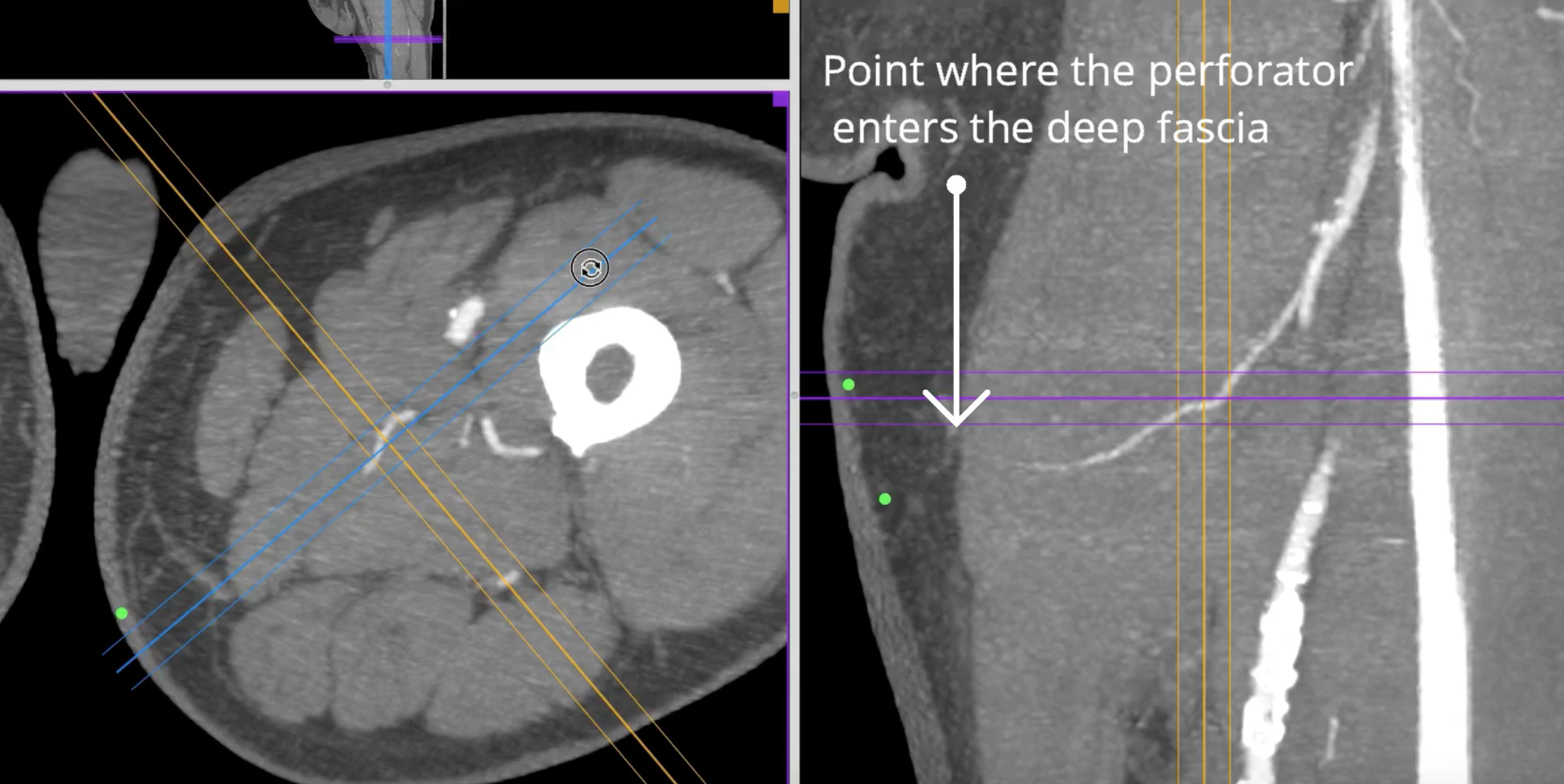
Figure 1. Computed tomographic angiography showing the point where the perforator enters the deep fascia, and it is divided in two branches within the subcutaneous tissue.
Morphology of the perforator and its branches, pedicle length and vertical location of the two branches from the ischial tuberosity can be assessed. The horizontal distance to the medial border of the gracilis muscle can be also measured, though it is not shown in the video. These measurements are translated on to the patient. Locations of both branches are confirmed with a handheld doppler (Figure 2).
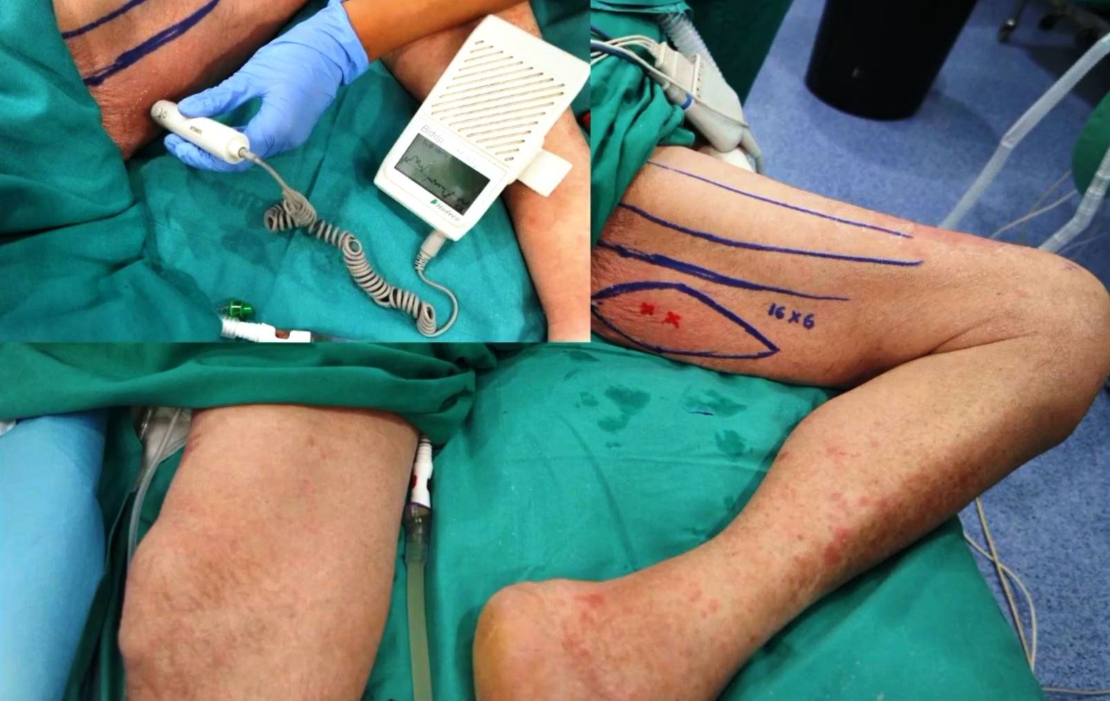
Figure 2. The measurements associated with the perforator and its branches are translated on to the patient. Locations of both branches are confirmed with a handheld doppler.
Flap elevation begins with an incision in the anterior margin of the flap to the superficial fascia layer, preserving the superficial vein. When the perforator is reached, dissection is continued to the deep fascia through the adductor magnus muscle until an adequate length is achieved. In this clinical case, the fat tissue between the two branches of the perforator is maintained.
Preoperative CTA is a useful technique to study location of perforators. This technique provides detailed anatomic information on morphology of perforators, course through the septum or muscle above the deep fascia and skin thickness. Preoperative 3-dimensional design of the flap can be done based on the characteristics of a selected perforator. Customization of the flap thickness is possible by modifying the plane of dissection.
CTA allows quick and easy assessment of the whole vascular anatomy of the leg and helps to arrive at the decision about selection of the best flaps based on the characteristics of the defect and on the individual anatomy of the patient. CTA suffers from the limitations of use of contrast medium and exposure of radiation to the patient. It may also lead to inadequate findings related to insufficient contrast enhancement which does not distinguish between the thin fat layer leading to a confusion between arteries and veins. Some of these limitations can be overcome by a color doppler ultrasound because it provides useful complementary information.
The thigh is a common donor site for soft tissue perforator flaps required for head and neck reconstruction because it can comfortably be accomplished by a two-teams approach. Among these, the anterolateral free flap is one of the most popularly used flaps for head and neck reconstruction and also for the anteromedial free flap. The profunda femoral artery perforator has been proposed recently as a good source for perforator flaps in head and neck reconstruction.
Received date: August 21, 2019
Accepted date: September 23, 2019
Published date: September 27, 2021
The study is in accordance with the ethical standards of the 1964 Helsinki declaration and its later amendments or comparable ethical standards.
The study did not receive any specific grant from funding agencies in the public, commercial, or not-for-profit sectors.
The authors report no financial or other conflict of interest relevant to this article, which is the intellectual property of the authors.
© 2021 The Authors. This is an open-access article distributed under the terms of the Creative Commons Attribution 4.0 International License (CC-BY).
Video A thin profunda artery perforator flap harvested from the left thigh is shown in this video. Preoperative computed tomographic angiography is used to assess morphology of the perforators and its branches, pedicle length and vertical location of the two branches from the ischial tuberosity. These measurements are translated on to the patient. Locations of both branches are confirmed with a handheld doppler. In conclusion, preoperative computed tomographic angiography is a useful technique to provide detailed anatomic information on morphology of perforators, course through the septum or muscle above the deep fascia and skin thickness. Computed tomographic angiography allows quick and easy assessment of the whole vascular anatomy of the leg and helps to arrive at the decision about selection of the best flaps based on the characteristics of the defect and on the individual anatomy of the patient.


The ALT and AMT flaps are the most commonly used thigh free flaps for intraoral reconstruction. Recently, PAP flap has been proposed as an alternative. This study aimed to compare the thickness of these thigh flaps and to identify the factors influencing flap thickness in our population.
Heredero S, Falguera-Uceda MI, Sanjuan A. The thin profunda artery perforator flap: Customized design and harvest. Int Microsurg J 2021;5(1):5. https://doi.org/10.24983/scitemed.imj.2021.00146
 PDF
PDF