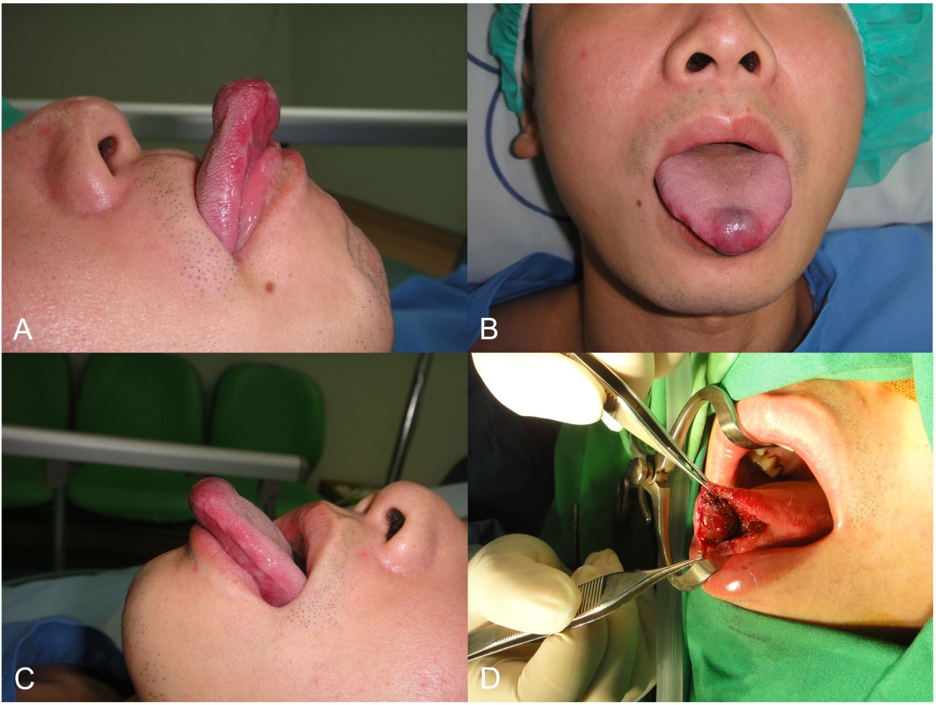
A 31-year-old man was presented for assessment of a mass in the midline of the ventral surface of the anterior tongue. The size of the lesion had varied since it was first noticed 3 months ago. According to his medical history, the onset of the mass was associated with trauma to the anterior tongue in a car accident. Examination of the tongue revealed a nontender, fluctuant, bluish mass of approximately 8 mm in diameter (Panel A-C). It was diagnosed that the mass was a mucocele derived from the glands of Blandin and Nuhn. We performed complete surgical excision of the mass (Panel D) and no recurrence had been detected during one year of follow-up.

Figure 1. Mucocele of the Blandin-Nuhn glands
The glands of Blandin and Nuhn are mainly mucus-secreting glands that are embedded within the muscle tissues of the anterior tongue ventrum [1]. The cystic formation of the Blandin-Nuhn glands on the ventral surface of the tongue is usually exophytic and may resemble diseases like pyogenic granulomata, polyps, or squamous papillomata [1]. The disease is an uncommon benign lesion, which is rarely found in adults [2]. Most of the cases reported in the medical literature were females [1]. The localization of lesion, a trauma history, rapid onset, variation in size, bluish color and fluid-filled consistency are helpful clues in the diagnosis of a mucocele derived from the glands of Blandin and Nuhn. Complete extirpation of the mucocele may be guaranteed by performing surgical excision of the lesion along with the associated glandular components to avoid recurrences. This is because the Blandin-Nuhn glands are not encapsulated, but are embedded to the musculature of the anterior tongue ventrum [3].
Received date: May 01, 2017
Accepted date: May 14, 2017
Published date: May 14, 2017
© 2017 The Author(s). This is an open-access article distributed under the terms of the Creative Commons Attribution 4.0 International License (CC-BY).
Kuo CL. Mucocele of the Blandin-Nuhn glands. Arch Otorhinolaryngol Head Neck Surg 2017;1(1):3. doi:10.24983/scitemed.aohns.2017.00011