International Microsurgery Journal (IMJ) is an open access journal with a comprehensive peer review policy and a rapid publication process. IMJ provides a global platform for scientists, clinicians, researchers and scholars to interact and share their latest research in all fields of microsurgery, including head and neck reconstruction, breast reconstruction, trunk and torso reconstruction, hand and upper extremities reconstruction, lower extremities reconstruction, lymphatic surgery, peripheral nerve, vascularized composite tissue allotransplantation, experimental microsurgery, microsurgical education, or other related topics.
IMJ is the official journal of International Microsurgery Club (IMC) and Internationl Course on Supermicrosurgery (国际超级显微外科学会, ICSM). International Microsurgery Club was created to facilitate discussion of complex cases and share knowledge of microsurgical techniques amongst experts around the world. Since its inauguration in May 2016, IMC now counts 5000 members and is one of the largest online educational platforms for microsurgery around the world. IMJ aims to make these educational case discussions available in written format to maximize the scholarly outreach and access to these valuable resources. IMJ welcomes submission of case reports, original articles, review papers, clinical studies, editorials, expert opinion and perspective articles, commentaries, and book reviews.
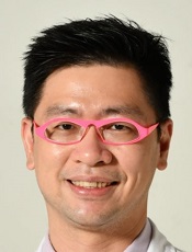
Dr. Tommy Nai-Jen Chang is a board-certified plastic surgeon in Taiwan. Currently, he is an Associate Professor in the Division of Reconstructive Microsurgery, Department of Plastic and Reconstructive Microsurgery, Chang-Gung Memorial Hospital, Linkou Medical Center, Taiwan. Dr. Chang finished his residency at Chang-Gung Memorial Hospital in 2011 and was appointed as staff of the department. He learned peripheral nerve reconstruction from Prof. David Chwei-Chin Chuang since 2012. Between 2015 and 2016, Dr. Chang completed a peripheral nerve research fellowship at the Hospital for Sick Children (SickKids) in Toronto, Canada, supervised by Dr. Gregory Borschel, Dr. Tessa Gordon and Dr. Ronald Zuker. After several years of peripheral nerve training under the guidance of Dr. David Chuang and Dr. Gregory Borschel, Dr. Chang is now capable of performing all types of peripheral nerve surgery. Currently, Dr. Chang works with Professor David Chwei-Chin Chuang and Dr. Johnny Chuieng-Yi Lu for peripheral nerve reconstruction for adult and pediatric brachial plexus injury, facial paralysis, and reconstruction of upper and lower extremity defects, decompression neuropathy including carpal tunnel, cubital tunnel, thoracic outlet syndrome, and the neurogenic tumors.
In addition to exploring motor nerves, Dr. Chang has also explored the sensory and autonomic nervous system as well in recent years and advanced some new areas, such as breast and nipple neurotization, robotic-assisted sympathetic trunk reconstruction, and hyper-selective neurectomy to treat primary hyperhidrosis. Besides, Dr. Chang also does microvascular surgery for Head and Neck reconstruction, perineal reconstruction, and extremities reconstruction. A total of 80 articles have been published in literature until the year 2022, mainly in the field of reconstructive microsurgery.
His research interests include peripheral nerve regeneration and reconstruction, and social media for microsurgery education. Dr. Chang founded the International Microsurgery Club (IMC) in 2016; until now, this group represents the largest online microsurgery platform. Dr. Chang was invited as the editor-in-chief of International Microsurgery Journal (IMJ) in 2017 and established International Microsurgery Website (IMW) in 2018 to provide educational resources for microsurgery surgeons worldwide. Dr. Chang coordinates the IMC Webinar Series during the COVID-19 pandemic to provide continuous education to global microsurgeons in 2020. By April 2022, there had been over 210 events, and all of the recordings were available on the IMW for members to access for free.
Aside from IMC, in Sep 2020, Dr. Chang also coordinated the main microsurgery webinar hosts and created an International Microsurgery Webinar League. Over 500 free accessed microsurgery-related free webinar talks were then connected online automatically, and the power of online learning of this system continues to expand and benefit the global microsurgeons.
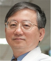
Prof. David Chwei-Chin Chuang is the first plastic surgeon who concentrated on peripheral nerve science that incorporated injuries, reconstruction, and research in Taiwan. It took him 30 years to be recognized as a world expert in the field of peripheral nerve injury and reconstruction. He started his microsurgical career in 1984 and became an academic professor in 2000. His phenomenal career was inspired largely by mentors namely: Dr. Samuel Noordhoff who motivated him to become a plastic surgeon, Dr. Fu-Chan Wei who influenced him to become a micro-surgeon; Dr. Julia K Terzis (USA), Dr. H. Millesi (Austria), Dr. A. Narakas (Switzerland), and Dr. Toru Kondo (Japan) - all four of whom had primed him to become a peripheral nerve microsurgeon.
In the process of aiming to make a solid contribution in the world of microsurgery, Prof. Chuang has developed many concepts and techniques related to the following: peripheral nerve reconstruction that deals with the classification level of brachial plexus injury; classification of level of radial nerve injury; classification of severity of post-paralysis facial synkinesis and strategy for treatment; the classification of traction avulsion amputation of the major limb; the technique of gracilis myocutaneous flap harvest; the technique revolution of FFMT for facial nerve reconstruction; the surgical treatment of thoracic outlet syndrome; the subfascial anterior transfer for cubital tunnel syndrome; the operative methods of nerve transfer for irreparable brachial plexus avulsion injury; the contralateral C7 transfer with free vascularized ulnar nerve graft; the intercostals nerve transfer to the musculocutaneous nerve, and other nerve transfer procedures.
Despite his already quite hectic schedule, he still found the time to get involved in many types of researches, which are related to peripheral nerve and free functioning muscle transplantation. Also, his numerous experimental types of research have been published in Plastic and Reconstructive Surgery, one of which is the research for arterialized venous flaps using vaso vasorum changes that he completed in 2015.
Because of his unique field of medical expertise, Prof. Chuang has been invited in countless local and worldwide meetings for special lectures, symposiums and panels in Japan, Korea, China, Thailand, Singapore, Indonesia, India, Argentina, Italy, Finland, Kuwait, Hong Kong, Mexico, Turkey, Germany and the USA. Furthermore, he also co-authored a chapter in three versions of the textbook entitled “Plastic Surgery” (2nd version 2006, third version 2013, and a new coming fourth version). Aside from these seemingly untiring achievements, he did several presentations locally and internationally, which were profiled in quite a number of publications for different medical journals.
Prof. Chuang has been similarly sought as the International Associate Editor of Plastic and Reconstructive Surgery from 2014 to 2016, and his term has been extended from 2016 to 2018. In relation to the foregoing, Prof. Chuang has twice hosted the "Instructional Course" for adult brachial plexus injury in 2009, and facial paralysis reconstruction in 2011. The third part (adult brachial plexus injury) of which, is coming soon this 2017.
To make his career in micro surgery more fulfilling, Prof. Chuang engaged himself with the Fellow Training Program of Chang Gung Memorial Hospital as one of the teachers, for hundreds of international foreign fellows. Along with this, he has treated international patients (approximately 100) from 20 different countries around the world.
As an authority figure in the field of micro-surgery, Prof. Chuang held vital positions as follows: Chief of Department of Plastic Surgery, Chang Gung Memorial Hospital, Taipei-Linkou; 13th President of Taiwan Plastic Surgical Association (2006-2008); 10th President of Taiwan Society for Surgery of the Hand (2009-2010); and First (1st) President of Taiwan Society for Reconstructive Microsurgery (2014-2016). At present, he is the 9th President of World Society for Reconstructive Microsurgery (WSRM) from March 2015 to June 2017. Prof. Chuang is undertaking every workable means and ways to fortify the relationship between the WSRM central office and the four regional societies (ASRM, ALAM, EFSM and APFSRM).
Prof. Chuang’s high hope is for the International Microsurgery Club and the International Microsurgery Journal to truly become the “World Society.”
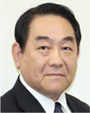
Professor Koushima, president of the World Society for Reconstructive Microsurgery from 2017 to 2019, graduated from Okayama Prefectural Tsuyama High School and Tottori University Faculty of Medicine in the 1970s, and completed his residency training in plastic surgery at Tokyo Women's Medical University Surgery Department and The University of Tokyo Hospital Plastic Surgery Department.
In 1983, he joined the University of Tsukuba Plastic Surgery Faculty of Medicine as a lecturer. By the year of 1990, he became an associate professor at Kawasaki Medical School Plastic Surgery. He was then sent to Harvard University for further education in 1996 and became a professor at Okayama University Faculty of Medicine Plastic Reconstruction Surgery, The University of Tokyo Plastic Reconstruction Surgery, and National University of Singapore in the early 2000s. In 2010, he was the associate dean at The University of Tokyo Hospital and became emeritus professor at The University of Tokyo Hospital as well as distinguished professor at Hiroshima University International Lymphedema Treating Center in 2017.
Throughout his academic career, he performed live surgery in 22 hospitals through invitations from international societies since 1997, including Ghent University Hospital, Belgium (twice), Louisiana State University, USA , Kuwait University hospital, München , Universidade de São Paulo hospital, Sant Pau Hospital, Barcelona, Ankara Medicine University, Turkey, China-Japan Friendship Hospital, Beijing (twice), National University of Singapore, Planas Hospital, Barcelona, Coimbatore city hospital, India, Versailles, France (2009/Feb), Utrecht University Hospital, Nederland (2009/May), Tomsk State University, Russia (2009/Sep), Planas Hospital, Barcelona(2010/Mar), University of Picardie Jules Verne, France (2010/Jul), Mahidol University, Thailand (2010/Aug), Hospital General Dr. Manuel Gea González (2010/Oct), The Chinese University of Hong Kong( 2010/Nov), National University of Singapore (2010/Dec), and Mumbai, India (2011/Mar).
Prof. Koushima’s contribution to the field can be divided into basic medical sciences and clinical medical sciences. He specializes in neuro-degeneration and neuro-regeneration, nerve grafting, tendon transplantation, and vascular pedicle nerve grafting. He is very well known in multiple clinical fields, such as perforator flap reconstruction, microvascular anastomosis, head and neck reconstruction, facial nerve palsy reconstruction, extremities’ reconstruction, lymphedema surgeries, as well as transgender surgeries for those with gender identity disorders. He has performed penis reconstruction, hand reconstruction, breast reconstruction, and aesthetic plastic surgery.
Prof. Koushima is affiliated with many societies as he is the reviewer of Plastic & Reconstructive Surgery Journal of American Society of Plastic Surgeons and Japanese Society of Plastic Reconstruction Surgery, and the founder and operator of International perforator flap conference 1997, Asia Pacific super micro conference in Singapore 2007, and Europe super micro conference in Barcelona 2010. He was the chairman of the Japanese Society for Surgery of the Hand, Japanese Society of Reconstructive Microsurgery, and Japanese Society for Lymphoreticular Tissue. He is the supervisor of the Japanese Society of Tissue Transplantation and members of Japan Society for Head and Neck Surgery and Japanese Research Society of Clinical Anatomy.
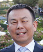
Prof. Wang is the world-famous microsurgeon in China. He is currently the chief of Shandong Provincial Hospital Extremities Surgery Department, professor and chief physician of Chinese Association of Microsurgery, vice chairman of Chinese Medical Association Branch of Microsurgery and Branch of Hand Surgery, and committee member of Shandong Province Society for Surgery of the Hand, committee member of Chief Shandong Province Society for Trauma surgery, vice chief physician of Shandong Province Society for Reconstruction Surgery, and editorial board member of Chinese Journal of Clinical Anatomy.
Prof. Wang’s work is filled with creativity, mainly focusing on a combination of clinical research and basic medical research. He has published several world-class original results based on innovative techniques in the clinical medical field. For instance, Dr. Wang’s research made great improvements regarding low temperature organ preservation. He successfully replanted three cases of low temperature-preserved severed finger at -196°C. Moreover, he created assembled digit reconstruction, a procedure that brought to light a whole new vision of the traditional procedure which has been used for thirty years in the world by replanting digits through cutting off the index toe. This new technique allows reconstructed fingers to resemble original digits instead of toes. His creativity and innovative visionary leads the field of reconstructive surgery into a new era.
Moreover, Prof. Wang excelled above and beyond expectations by taking the time to review and revise microsurgery anatomy, which clarified certain anatomy structures associated with microsurgery. As a result of his contributions, reconstructive microsurgery has been significantly improved. Furthermore, he has written many literary works pertaining to such topics, such as the "Clinical Anatomy Atlas of Microsurgery".
It is indeed a significant step forward in the development of global microsurgery to have Prof. Wang serve as the honorary Editor-in-Chief of the International Microsurgery Journal since with this collaboration, we have succeeded in removing the barriers of language and platform, so people are able to learn the techniques from Chinese microsurgeons. There will be more cooperation not only in the journal, but also in the conference and the online platform in the future. This knowledge exchange and pooling should be beneficial to global microsurgeons and patients alike.

Dr. Peter Neligan is Irish and attended Trinity College Dublin, graduating in 1975. He obtained a General Surgery Fellowship in 1980 then Plastic Surgery training in Ireland. Moving to Toronto in 1983 and completing fellowships in research, pediatric plastic surgery, microvascular surgery and burn surgery. He received FRCSC in Plastic Surgery in 1988. He became Chair of Plastic Surgery at the University of Toronto in 1996 and Wharton Chair in Reconstructive Plastic Surgery in 1999. In 2007 he moved to Seattle as Professor of Surgery and Director of the Center for Reconstructive Surgery at the University of Washington. His practice is in microsurgery, perforator flaps, lymphatic surgery, facial re-animation and VCA. He was retired in 2020.
Publications include more than 12 books, 85 book chapters and over 200 peer-reviewed papers. He is the Editor-in-Chief of Plastic Surgery, a 6 Volume textbook which has become the standard text for Plastic Surgery Training Programs around the world. He’s been invited to over 300 universities or major societies as a visiting professor or honored guest. He sits on several editorial boards and is past Editor-in-Chief of the Journal of Reconstructive Microsurgery.
Dr. Neligan is Past-President of the PSF, ASRM as well as the NASBS. He is also a past board member of the AHNS. He is a past trustee of the ASPS. He is married to Gabrielle Kane, a radiation oncologist and they have 2 adult children and 4 grandchildren.
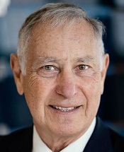
Dr. Ronald M. Zuker, graduated from the Faculty of Medicine at the University of Toronto and after completing his Plastic Surgery training under Dr. W. K. Lindsay, embarked on a McLaughlin Travelling Fellowship learning about microsurgery, cleft care and burns in Japan, Australia, New Zealand and Europe. Dr. Zuker was appointed to the staff at the Hospital for Sick Children in 1978 where he has remained and continues to be active to this day. He was Chief of the Division of Plastic & Reconstructive Surgery from 1986 to 2002.
Surprisingly Dr. Zuker began his medical career in the Amazon Basin as a river doctor working for the Hospital Amazonica Albert Schweitzer in Pucullpa, Peru where his fluency in Spanish paid dividends. To this day, his commitment to global health continues with strong representation in Operation Smile. After he completed his surgical training, he was integral in establishing the microsurgery program at the University of Toronto with Drs. Ralph Manktelow, Nancy McKee, Howard Clarke and Jim Mahoney. Quickly, Toronto became known as an epicenter of excellence and attracted dozens of trainees from around the globe including Professor Fu Chan Wei who, stimulated by his teachers, formed the world’s largest microsurgery unit at Chang Gung Hospital in Taipei.
Dr. Zuker is one of the original pioneers of microsurgery. He is a founding member of the American Society of Reconstructive Microsurgery and the Group for Advancement of Microsurgery (GAM – Canada). He has had key leadership positions in our specialty’s important societies including the American Academy of Pediatrics, the American Association of Plastic Surgeons, the American Association of Pediatric Plastic Surgeons, the American Burn Association, the American Cleft-Craniofacial Association, the American Society for Reconstructive Microsurgery, the American Society of Peripheral Nerve, the American Society of Plastic Surgeons, the Congress of the International Microsurgery Society, the Canadian Society of Plastic Surgeons amongst many others, culminating with the Presidency of the International Society of Plastic Reconstructive and Aesthetic Surgery Congress in Vancouver 2011.
His curriculum vitae is rich with accolades, honors, peer-review papers and scientific presentations. He has been teaching residents and medical students in Plastic Surgery since 1978. He has taught fellows from all over the world. He has given lectures on 4 of the 5 major continents. He has published more than 100 peer-reviewed scientific papers and book chapters. One of his most significant contributions has been his textbook entitled Principles and Practice of Pediatric Plastic Surgery (co-edited with Bruce Bauer and Mike Bentz) which has become the go-to book for Pediatric Plastic and Reconstructive Surgery and is heading for a 3rd edition any day now! He has acted as a great mentor for the latest generation of reconstructive microsurgeons and is generous in sharing his surgical secrets.
It is easy to say that the backbone of Dr. Zuker’s career has been his dedication to clinical excellence and innovation. The Zuker name has been integral in shaping the sub-specialty of pediatric plastic surgery through his contributions to microsurgery, cleft lip and palate and upper extremity surgery. However, Dr. Zuker will be best known for his work as the “Smile Doctor”. His passion has been the restoration of facial function in patients with facial paralysis and Moebius syndrome. To this end, he has revolutionized the approach to the management of established facial paralysis using free functioning muscle transfer with the gracilis muscle being the workhorse. With Dr. Ralph Manktelow, he developed innovative and novel techniques for transfer of the gracilis muscle from the leg to restore the smile in patients with facial paralysis. Surgeons from around the world visit him to learn this remarkable procedure. He has been recognized for his passion by the Moebius Foundation and the Smile Foundation of South Africa. There is a photo in the collage of Ron’s office memorabilia showing him shaking hands with Nelson Mandela in recognition of his service to patients with facial paralysis. Ron holds honorary degrees as Fellow of the Royal College of Surgeons of Edinburgh and Honorary Fellowship of the College of Plastic Surgeons of South Africa.
Everyone is well aware of Dr. Zuker’s interest in smile surgery but here are ten rarely known facts about Dr. Ron Zuker’s clinical achievements:
Dr. Zuker’s career began with a focus on the delivery of health care to those in greatest need. He continues to exhibit this high level of altruism by devoting a huge amount of his energy to helping children with clefts in low and middle-income countries spending time in South America, Africa and India with the Operation Smile group. His latest endeavor is the creation of the Pediatric Vascularized Composite Allotransplantation program at the Hospital for Sick Children pushing the clinical limits of reconstructive surgery.
It is a small world under the microscope, but it is incredible how far it can take you in the real one. His name is synonymous with excellence and innovation. In the arena of innovative reconstructive microsurgery, Ron has few equals. Dr. Ron Zuker has made significant innovations in the unique areas of conjoined twins, composite vascularized allotransplantation, free functioning muscle transfer, solid organ transplantation and global outreach. In the field of Plastic & Reconstructive Surgery, Ron is an icon and for his contributions to the specialty was awarded the Canadian Society Lifetime Achievement Award in June 2014.

Dr. Gregory Buncke is a plastic, hand, and microsurgeon. Currently, he is the director of the Buncke Clinic in San Francisco, and he is the son of the pioneering microsurgeon Harry J. Buncke. Dr. Buncke earned his medical degree from Georgetown University in Washington, DC. He completed his residency in Plastic and Reconstructive Surgery at Stanford University and did a fellowship in Hand and Microsurgery at the Davies Medical Center in San Francisco, where he remains in practice. Dr. Buncke is Board Certified by the American Board of Plastic Surgery and has a Certificate of Added Qualification in Surgery of the Hand. He is a Fellow of the American College of Surgeons, and a member of several societies, including the American Society of Plastic Surgeons, the American Society for Reconstructive Microsurgery, and the American Society for Surgery of the Hand. Dr. Buncke has published numerous articles related to a wide variety of plastic, hand, and microsurgery topics.
Since 1970 The Buncke Clinic has been at the forefront of the advancement of reconstructive surgery. The Buncke Clinic is not only known for its rich history, but also contributes to the advancement of microsurgery in numerous ways, including outstanding clinic service, research, and educational programs. Its customized website also provides all the essential background knowledge for microsurgeons and the updated master talk organized by Dr. Bauback Safa. You can find out more about the Buncke Clinic online, as well as all the secrets about microsurgery, by visiting their websites.
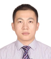
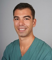
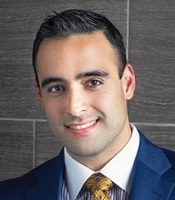
Dr. Matteo Amoroso, is a consultant at the department of Plastic and Reconstructive Surgery, Karolinska University Hospital. His clinical interest focus on reconstructive microsurgery of the breast, head and neck, lower and upper extremities. Since 2015, he is a board certified Plastic Surgeon in both Sweden and Italy where he completed his Plastic and Reconstructive Surgery residency training (2009-2015). He received further surgical training working in 2014-2015 with Prof. Ömer Özkan focusing on experimental microsurgery , head and neck reconstruction including the face transplant patients, operated at the Akdeniz University Hospital, Antalya, Turkey. In 2017, he moved in Taiwan for a one-year Clinical Fellowship in Microsurgery at the China Medical University Hospital directed by Professor Hung-Chi Chen. Dr. Amoroso´s research area focus physiology and hemodynamic in perforator flaps and as well as in tissue engineering. Indeed, he has an active collaboration with Chalmers University of Technology in Sweden, on 3D Bioprinting of human cartilage. He is member of the Swedish societies of Plastic Surgery (SPKF) and Microsurgery (SFRM), author of several book chapters and articles published on international journals.
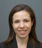
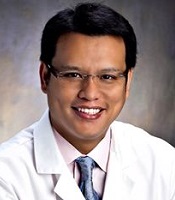

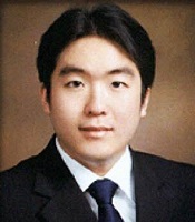
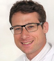
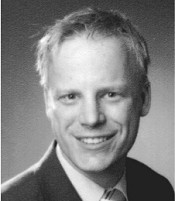
Dr. Eisenhardt is a Professor of Plastic Surgery at the University of Freiburg/Germany. He graduated from Freiburg University in 2005 and after his postdoctoral studies in Melbourne/Australia completed his Plastic surgery and microsurgery training at the University of Freiburg Medical Centre. He is specializing in complex reconstructive microsurgery, particularly facial paralysis reconstruction and extremity reconstruction. Dr. Eisenhardt has a strong passion for student and resident training and he is responsible for the Microsurgery training program at his institution. Dr. Eisenhardt is PI of several major grants of the German Research Foundation (DFG) investigating multiple aspects of microsurgery and microcirculation and was awarded a Heisenberg Fellowship in 2014, the most prestigious personal fellowship awarded by the DFG, honoring his scientific contribution to the field of microcirculation research.
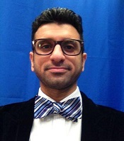
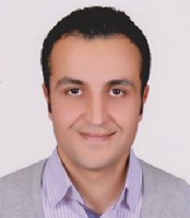
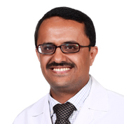
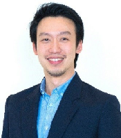
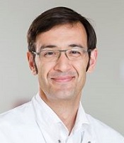
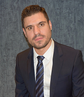
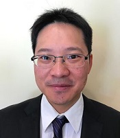
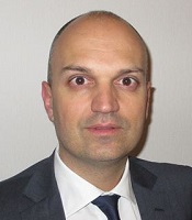
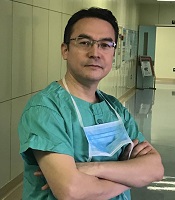
Dr. Andrés Rodríguez Lorenzo, after plastic surgery residency in Spain, completed his two-year microvascular fellowship at the Cannieburn Plastic Surgery Unit in Glasgow and Chang Gung Memorial Hospital in Taiwan. He is Associate Professor for Plastic Surgery and Chief of Plastic Surgery at Uppsala University Hospital, leading one of the most active microsurgical centers in Scandinavia. He is the program director of the International Fellowship Program in Microsurgery at the Uppsala University and his main focus of research is head and neck reconstruction including preclinical studies on face transplantation, virtual planning and facial paralysis.
This study introduces an advanced tubularized radial artery forearm flap (RAFF) technique, marking an enhancement over traditional methods in addressing complex nasal reconstructions. It integrates functional and aesthetic considerations through a structured, multi-stage reconstruction process, emphasizing the use of tubularized flaps. Key learning points include the detailed crafting of stable nasal passages, strategic use of costal cartilage for robust structural support, and tailored postoperative care with silicone splints. The tubularized RAFF technique not only optimizes patient outcomes and quality of life but also provides plastic surgeons with critical insights to refine their techniques in facial reconstruction. Indispensable for professionals in the field, this article enriches the understanding of sophisticated reconstructive challenges and solutions.
This manuscript showcases an advanced surgical approach for treating malignant giant cell tumor of bone, emphasizing precision and ethical considerations. It leverages innovative pedicled flap technologies, as opposed to free flaps, enhancing limb functionality and patient quality of life. This technique equips surgeons with evidence that tailored surgical strategies can significantly improve outcomes in complex cases. The paper discusses technical challenges and highlights the application of supercharging and superdrainage techniques in limb reconstructions, methods well-established in microsurgery but infrequently used in oncological contexts. These techniques are crucial for optimizing flap viability and ensuring surgical success. Additionally, the manuscript underscores the profound impact of these advancements on patient lives, offering hope and showcasing tangible benefits. This narrative, blending scientific analysis with patient stories, enriches the understanding of limb reconstruction innovations in oncological surgery, making it invaluable for surgeons.
The groundbreaking study illuminates the complex mechanisms of nerve regeneration within fasciocutaneous flaps through meticulous neurohistological evaluation, setting a new benchmark in experimental microsurgery. It challenges existing paradigms by demonstrating the transformative potential of sensory neurorrhaphy in animal models, suggesting possible clinical applications. The data reveal a dynamic interplay of nerve recovery and degeneration, offering critical insights that could revolutionize trauma management and reconstructive techniques. By bridging experimental findings with hypothetical clinical scenarios, this article inspires continued innovation and research, aimed at enhancing the efficacy of flap surgeries in restoring function and sensation, thus profoundly impacting future therapeutic strategies.
Our belief is: “Stronger microsurgeons for a better world.”
The progress of microsurgery in developing low-income countries.
Professor David Chwei-Chin Chuang sincerely welcomes you to join them for this instructional course on Nov. 13-16, 2017 at Chang-Gung Memorial Hospital.
A novel technique of sequential ETS micro-venous anastomoses using three vessel loops for IJV occlusion and a single vascular clamp to retract and hold the anastomoses sites in position.
A personal experience in robotic microsurgery.
A Fantastic platform for learning, professional development, and camaraderie. It's all just a few clicks away.
Division of the lateral plantar artery does not jeopardize the foot circulation because of anastomosis of the lateral plantar artery with the dorsalis pedis artery at the first intermetatarsal space. However, care should be taken with patients with peripheral artery occlusive disease and the flow of dorsalis pedis artery should be confirmed before surgery. Given the advantages of sizable vessel, easy dissection, and proximity to the defect, we believe that the lateral plantar artery might be a valuable option as recipient vessel for lateral plantar forefoot reconstruction.
Health care systems in many countries are confronted with increasing economic limitations. Thus, complex microsurgical procedures and extensive rehabilitation programs are poorly compensated. However, this case demonstrates a dramatic reduction of socioeconomic expenses by allowing a potential radiocarpal amputee to return to work for another estimated 30 years.
The 2nd instructional course for adult brachial plexus injuries is a fantastic opportunity to meet and learn from experts in the field. We cordially invite you to visit Chang Gung Memorial Hospital to participate in this unique learning experience.
Pedicled anterolateral thigh flap is a versatile option for reconstruction of complex soft tissue defects in varied anatomical regions. Its wide arc of rotation and less donor site morbidity are its added advantages.
The authors reviewed the MDCT images to show the number of lymph nodes superior to the saphenofemoral junction. In this study, on average, 3.67 nodes existed. However, there were 4 percent of cases with no countable nodes. This result indicates that appropriate preoperative screening is needed for this procedure.
The communication among international microsurgeons have switched from one direction (from paper, textbook) to multiway interactions through the internet. The authors believe the online platform will play an immensely important role in the learning and development in the field of microsurgery.
A 29-year-old male suffered from total loss of function of his left shoulder and upper limb after a traffic accident while riding a motorcycle. He was treated by a brachial plexus specialist at another hospital and managed surgically under the impression of total root avulsion.
The authors present a novel synthetic vascular model for microanastomosis training. This model is suitable for trainees with intermediate level of microsurgical skills, and useful as a bridging model between simple suturing exercise and in vivo rat vessel anastomosis during pre-clinical training.
Combining intraoperative ICG lymphography with NIR vein visualization can aid supermicrosurgeons in identifying lymphatic vessels and superficial venules to guide LVA incision placement. This guided approach significantly improves successful creation of LVAs when compared to the blind (anatomic) approach. In addition, the absence of linear ICG lymphographic patterns does not prevent formation of successful LVAs.
Authors present a case of combined proximal median and ulnar nerves extensive injury that was successfully managed using a novel safe strategy implemented.
Authors report a case of lower extremity lymphedema treated by LVA that preoperatively mapped not only lymphatic vessels by PDE, but also veins and venules using Veinsite™ .
Shoulder instability and lack of shoulder mobility are common and exhausting problems in adult patients with traumatic brachial plexus injury. Although primary nerve reconstruction remains the “gold standard” for brachial plexus management, secondary shoulder reconstruction by trapezius transfer is needed in neglected cases.
Immediate limb compression following the LVA procedure facilitates lymphatic drainage and increases the surgical efficacy by increasing the number of functioning anastomoses, and is a recommended postoperative practice.
Optimum operating conditions are an integral part of a successful microvascular surgery. We hereby introduce a new technique to make a low- cost microsuction cannula, which will be helpful for microvascular surgeons.
Improving efficiency of microvascular anastomosis by combination of open-loop and airborne suture techniques.
The authors describe various patient and breast-related factors that influence surgical outcomes while also addressing some techniques and principles for aesthetic microsurgical reconstruction.
Supraclavicular flap is an excellent fasciocutaneous flap for head and neck reconstruction due to its close color and texture match. In general, long flaps are required, but with the risk of distal necrosis. The aim of this study is to assess the relationship between the length and distal end necrosis of the supraclavicular flap.
This case report demonstrates an important supermicrosurgical technique for lymphedema, which was established by Isao Koshima in 1994. So far, over 2,000 cases of limb edema have been treated by this surgical method.
The authors present a revised application of the composite iliac crest bone free flap for hemimaxillectomy defects. This flap solely can give a support to the eye globe and the dental implants without osteotomies and titanium meshes. The upper ridge of the flap can be shaped to replicate the curvature of the orbital floor. The inner oblique muscle and the deep fascia can serve as an additional material for separation of the oral cavity from the nasal airway. Utilization of the Customized Contour Implants gives a surgeon a refining instrument for aesthetic correction of facial projection.
The authors proposed a new less invasive island flap, namely the first metatarsal artery capillary perforator flap. The advantages of this flap include the preservation of the first metatarsal artery and the adiposal tissue in the web space, thereby preventing compression around the remaining deep peroneal nerve.
The authors present a retrospective review of 7 patients who underwent wide excision of the malignant tumors around the clavicle. Patient demographics, clinical details, the arc of rotation, outcome, and complications were analyzed.
Patients with gynecological abdominal wall malignancies can benefit significantly from radical resection and autologous reconstruction. The pedicled anterolateral thigh flap is the preferred donor site, offering a reliable solution to abdominal wall reconstruction in this setting. The satisfactory results should prompt a more aggressive surgical approach for these patients. This article describes the authors' experiences with the abdominal reconstruction following surgical resection of gynecological abdominal wall malignancy using pedicled anterolateral thigh flap.
Prof. Koushima, president of World Society for Reconstructive Microsurgery, proposes an innovative concept and technique of the multi-stage ‘Orochi’ combined flaps (sequential flaps in parallel). The technique opens a new vista in reconstructive microsurgery.
Brachial plexus imaging is challenging due to its anatomical complexity. Multiple sequences of MRI may be performed pre-operatively for an accurate radiological diagnosis of brachial plexus injury. This image shows the useful role of MRI in localization of acute brachial plexus injury.
Traditionally, suturing techniques have been the mainstay for microvascular anastomoses, but owing to its technical difficulty and labour intensity, considerable work has gone into the development of sutureless microvascular anastomoses. In this review, the authors take a brief look at the developments of this technology through the years, with a focus on the more recent developments of laser-assisted vascular anastomoses, the unilink system, vascular closure staples, tissue adhesives, and magnets. Their working principles, with what has been found concerning their advantages and disadvantages are discussed.
Vessel-compromised neck is a challenge for the microvascular surgeon planning for a free tissue transfer. This study describes the authors’ experience regarding a successful free tissue transfer in a vessel-compromised neck. The authors also propose an algorithm for management of vessel-compromised neck.
The authors demonstrated the utility of hemi-intravascular stenting technique in a pediatric thumb replant.
Facial palsy affects both the functional and aesthetic aspects of the face. Many techniques for both static and dynamic facial reanimation have been described. Authors present their series of facial reanimation using the functioning free gracilis muscle flap innervated by the masseteric nerve.
This study reviews the reconstruction modalities within the multidisciplinary approach for chest wall reconstruction. The authors have proposed an algorithm that is formulated based on their experience to manage chest wall defects following cancer resection.
LVA and vascularized lymph node transfer VLNT are established lymphedema treatments. However, LVA is only effective for early disease and VLNT can cause donor-site lymphedema and contour deformity. VLVT is free of these limitations. The authors described their experience of a new VLVT technique.
The ALT and AMT flaps are the most commonly used thigh free flaps for intraoral reconstruction. Recently, PAP flap has been proposed as an alternative. This study aimed to compare the thickness of these thigh flaps and to identify the factors influencing flap thickness in our population.
Authors discuss a silicone tube that provides structural support to vessels throughout the entire precarious suturing process. This modification of the conventional microvascular anastomosis technique may facilitate initial skill acquisition using the rat model.
Authors proposed a simple innovative technique that helps in achieving a clear surgical field at the time of venous repair by diverting the venous bleeding through a glove finger and at the same time preventing venous congestion of the flap.
PEDs can be used as alternative means of magnification in microsurgery training considering that they are superior to surgical loupes in magnification, FOV and WD ranges, allowing greater operational versatility in microsurgical maneuvers, its behavior being closer to that of surgical microscopes in some optical characteristics. These devices have a lower cost than microscopes and some brands of surgical loupes, greater accessibility in the market and innovation plasticity through technological and physical applications and accessories with respect to classical magnification devices. Although PEDs own advanced technological features such as high-quality cameras and electronic loupes applications to improve the visualizations, it is important to continue the development of better technological applications and accessories for microsurgical practice, and additionally, it is important to produce evidence of its application at surgery room.
Avulsion injuries and replantation of the upper arm are particularly challenging in the field of traumatic microsurgery. At present, the functional recovery of the avulsion injuries upper arm after the replantation is generally not ideal enough, and there is no guideline for the surgeries. The aim of this study was to analyze the causes of failure of the upper arm replantation for avulsion injuries, summarize the upper arm replantation’s indications, and improve the replantation methods.
VSG is a type of bariatric surgery that is accepted as the simplest but most efficient surgical modality to treat morbid obesity. A mouse model of VSG has been widely utilized to investigate the underlying mechanisms of VSG. There are numerous microsurgical techniques for VSG in mice, and we report herein a modified technique with 2 disposable micro-clamps to simplify the surgery.
This article presented 3 cases diagnosed with chronic fatigue syndrome, severe skin adjuvant infiltration and skin ulcers.
This case report describes authors successfully attempt to perform a topographically correct sural nerve transplantation in the extracranial facial nerve stem using the intraneural facial nerve stem topography proposed by Meissl in 1979. This is the first reported case of successful fascicular nerve grafting of the facial nerve stem following extensive laceration.
ICG lymphography is an invaluable tool in lymphedema management. Both immediate and delayed scans are needed when performing the study. The delayed scan needs to be performed at the time of the lymphographic plateau to appreciate the full extent of the pathology. Using a recumbent cross trainer, the lymphographic plateau can be achieved in 15 minutes following ICG injection. We have found this exercise enhanced ICG lymphography protocol worthwhile of adoption by high volume lymphedema centers to raise diagnostic accuracy and efficiency.
The implications of rebound heparin hypercoagulability following cessation of therapy in microsurgery is unreported. In this article the authors report two cases of late digit circulatory compromise shortly after withdrawal of heparin therapy. The authors also propose potential consideration for changes in perioperative anticoagulation practice to reduce this risk.
The supraclavicular flap has gained popularity in recent years as a reliable and easily harvested flap with occasional anatomical variations in the course of the pedicle. The study shows how the determination of the dominant pedicle may be aided with indocyanine green angiography. Additionally, the authors demonstrate how they convert a supraclavicular flap to a free flap if the dominant pedicle is unfavorable to a pedicled flap design.
A thin profunda artery perforator flap harvested from the left thigh is shown in this video. Preoperative computed tomographic angiography is used to assess morphology of the perforators and its branches, pedicle length and vertical location of the two branches from the ischial tuberosity. These measurements are translated on to the patient. Locations of both branches are confirmed with a handheld doppler. The authors concluded that preoperative computed tomographic angiography is a useful technique to provide detailed anatomic information on morphology of perforators, course through the septum or muscle above the deep fascia and skin thickness. Computed tomographic angiography allows quick and easy assessment of the whole vascular anatomy of the leg and helps to arrive at the decision about selection of the best flaps based on the characteristics of the defect and on the individual anatomy of the patient.
The senior author (Dr. Isao Koshima) designed a tibial osseo-periosteal (TOP) flap. TOP flap has a favorable anatomical position with a thin skin around it, hence it is a good option for an island flap. TOP flap can be used for various mild to moderately sized osteo-cutaneous defects with low morbidity. In this article, the authors describe their experience of the first reported cohort of TOP flaps in clinical practice.
The video presents a useful technique for microvascular anastomosis in reconstructive surgery of the head and neck. It is advantageous to use this series of sutures when working with limited space, weak vessels (vessels irradiated, or with atheroclastic plaques), suturing in tension, or suturing smaller vessels (less than 0.8 cm in diameter).
Osteoarthritic finger joints are often repaired with joint implants, arthrodesis, or a vascularized interphalangeal joint graft. However, grafts can damage the donor toe. Based on the results of this study, the authors suggest that vascularized distal interphalangeal joint transfers from the second toe may be suitable for reconstructing these defects through microsurgery.
The likelihood of donor site ischemia following the harvesting of a fibula flap is extremely low, but it is potentially lethal if it occurs. The authors describe a case of ischemia of the lower extremity following a free fibula harvest for head and neck reconstruction. The authors discuss preoperative, intraoperative, and postoperative strategies to assist in diagnosing and managing risks associated with free fibula flap harvesting in this paper.
In a cost-effective and portable way, a novel method was developed to assist trainees in spinal surgery to gain and develop microsurgery skills, which will increase self-confidence. Residents at a spine surgery center were assessed before and after training on the effectiveness of a simulation training model. The participants who used the training model completed the exercise in less than 22 minutes, but none could do it in less than 30 minutes previously. The research team created a comprehensive model to train junior surgeons advanced spine microsurgery skills. The article contains valuable information for readers.
The loupe plays a critical role in the microsurgeon's arsenal, helping to provide intricate details. In the absence of adequate subcutaneous fat, the prismatic lens of the spectacle model may exert enormous pressure on the delicate skin of the nasal bone. By developing a soft nasal support, the author has incorporated the principle of offloading into an elegant, simple yet brilliant innovation. A simple procedure such as this could prove invaluable for microsurgeons who suffer from nasal discoloration or pain as a result of prolonged use of prismatic loupes. With this technique, 42% of the pressure applied to the nose is reduced.
Smartphones and cellular technology have revolutionized flap monitoring. Smartphones provide low-cost thermal imaging alternatives to flap monitoring. It remains unclear, however, whether this method is accurate or reliable. It may be challenging to use smartphone thermal imaging when clinicians fail to communicate clinically relevant events. The authors demonstrate three instances in which the thermal imaging information on smartphones was misleading. Each case is analyzed to determine how clinical decisions should be made.
The article describes the case of a 27-year-old man who suffered from scarring at the penopubic junction as a result of penis lengthening surgery. The authors explain how they repaired the penopubic junction by using a pedicled superficial circumflex iliac artery perforator flap. The authors also propose a safe and effective hybrid technique for harvesting superficial circumflex iliac artery perforator flaps. According to the authors, the pedicled superficial circumflex iliac artery perforator flap is an effective method for reconstructing the penopubic scar.
An examination of plastic surgery residents' experiences with microsurgery in Latin American countries was conducted in a cross-sectional study with 129 microsurgeons. The project also identifies ways to increase the number of trained microsurgeons in the region. The authors claim that there are few resident plastic surgeons in Latin America who are capable of attaining the level of experience necessary to function as independent microsurgeons. It is believed that international microsurgical fellowships would be an effective strategy for improving the situation.
A significant increase in peripheral nerve surgery has occurred in recent years due to improvements in surgical techniques. In most reconstructive procedures, sensory restoration is frequently neglected in preference to restoring motor function. Along with increasing the risk of developing injuries to the body, patients who lose protective sensations are more likely to develop neuropathic pain and depression, which adversely affect their quality of life. As regaining sensory function is important, the study examines a variety of techniques that may be useful for restoring sensory function across various body parts.
This retrospective study on the keystone design perforator island flap (KDPIF) reconstruction offers valuable insights and compelling reasons for readers to engage with the article. By sharing clinical experience and reporting outcomes, the study provides evidence of the efficacy and safety profile of KDPIF as a reconstructive technique for soft tissue defects. The findings highlight the versatility, simplicity, and favorable outcomes associated with KDPIF, making it an essential read for plastic surgeons and researchers in the field. Surgeons worldwide have shown substantial interest in KDPIF, and this study contributes to the expanding knowledge base, reinforcing its clinical significance. Moreover, the study's comprehensive analysis of various parameters, including flap survival rate, complications, donor site morbidity, and scar assessment, enhances the understanding of the procedure's outcomes and potential benefits. The insights garnered from this research not only validate the widespread adoption of KDPIF but also provide valuable guidance for optimizing soft tissue reconstruction in diverse clinical scenarios. For readers seeking to explore innovative reconstructive techniques and improve patient outcomes, this article offers valuable knowledge and practical insights.
This comprehensive review article presents a profound exploration of critical facets within the realm of microsurgery, challenging existing paradigms. Through meticulous examination, the authors illuminate the intricate world of microangiosomes, dissection planes, and the clinical relevance of anatomical structures. Central to this discourse is an exhaustive comparative analysis of dermal plexus flaps, meticulously dissecting the viability and potential grafting applications of subdermal versus deep-dermal plexi. Augmenting this intellectual voyage are detailed illustrations, guiding readers through the intricate microanatomy underlying skin and adjacent tissues. This synthesis of knowledge not only redefines existing microsurgical principles but also opens new frontiers. By unearthing novel perspectives on microangiosomes and dissection planes and by offering a comparative insight into dermal plexus flaps, this work reshapes the landscape of microsurgery. These elucidations, coupled with visual aids, equip practitioners with invaluable insights for practical integration, promising to propel the field of microsurgery to unprecedented heights.
This article presents a groundbreaking surgical approach for treating facial paralysis, focusing on the combination of the pronator quadratus muscle (PQM) and the radial forearm flap (RFF). It addresses the challenges in restoring facial functions and skin closure in paralysis cases. The study's novelty lies in its detailed examination of the PQM's vascular anatomy when combined with the RFF, a topic previously unexplored. Through meticulous dissections, it provides crucial anatomical insights essential for enhancing facial reanimation surgeries, offering significant benefits in medical practices related to facial reconstruction and nerve transfer techniques.
This article explores the transformative impact of artificial intelligence on healthcare, providing readers with insights into its role in enhancing diagnostics, treatment, and patient care. It highlights potential cost savings and promotes a proactive approach to health management. The piece underscores advancements in robot-assisted surgery and artificial intelligence-enhanced virtual nursing, alongside efficient data management in healthcare. It addresses challenges and ethical considerations in integrating artificial intelligence, emphasizing the need to maintain clinical skills and empathy. Beneficial for readers, the article advocates a balanced approach that melds technological innovation with foundational healthcare principles, aiding informed decision-making in evolving healthcare landscapes.
This article unveils the hidden treasures within medical case reports, showcasing their historical significance, real-world relevance, and capacity to inspire innovation. By dissecting eight pivotal factors influencing the fate of case reports in contemporary academic publishing, it offers readers a profound understanding of the challenges and opportunities these narratives present. Readers will gain an enhanced appreciation for the essential role of case reports in linking clinical practice with research, highlighting rare conditions, new treatments, and unusual clinical cases. Furthermore, this article provides practical strategies for journals and the academic community to promote inclusivity, balance, and excellence in the publication of case reports, making it a must-read for anyone in the medical field.
This article holds critical relevance for healthcare professionals, particularly in the fields of microsurgery, oncology, and vascular medicine. It thoroughly examines the diagnostic challenges faced in distinguishing between recurrent lymphedema and deep vein thrombosis in elderly cancer patients following lymphovenous anastomosis surgery. It highlights the significant risk of misdiagnosing deep vein thrombosis as lymphedema, a mistake that can delay critical treatment due to their clinical similarities. The case study of a 79-year-old patient emphasizes the importance of a comprehensive reassessment, considering the patient's entire medical history, including the effects of cancer treatments like immunotherapy. The article stresses the need for a holistic approach to patient management and the utilization of advanced diagnostic tools for accurate diagnosis and treatment. It is essential reading for its insights into the complex dynamics of postoperative care and the critical importance of accurate diagnosis in treating elderly cancer patients effectively.
This article presents a case study on the successful replantation of a pediatric lower lip after a dog bite, focusing on artery-only microanastomosis. It highlights the challenges and effective strategies in pediatric microvascular surgery, particularly emphasizing the importance of specialized surgical techniques and thorough postoperative care. Alongside the case study, an extensive literature review supports the feasibility of artery-only anastomosis and the traditional yet critical use of leech therapy for managing venous congestion. This research is vital for medical professionals specializing in pediatric surgery, offering key insights into improving both functional and aesthetic outcomes for young patients. Additionally, it identifies gaps in long-term research and stresses the need for ongoing studies to refine treatment protocols, making it an indispensable resource for enhancing patient care and outcomes in pediatric reconstructive surgeries.
This article exemplifies a significant advancement in microsurgical techniques, highlighting the integration of robotic-assisted surgery into the deep inferior epigastric perforator (DIEP) flap procedure for breast reconstruction. It demonstrates how innovative robotic technology refines traditional methods, reducing the invasiveness of surgeries and potentially lessening postoperative complications like pain and herniation by minimizing the length of the fascial incision. This manuscript is pivotal for professionals in the medical field, especially those specializing in plastic surgery, as it provides a comprehensive overview of the operative techniques, benefits, and critical insights into successful implementation. Moreover, it underscores the importance of ongoing research and adaptation in surgical practices to enhance patient outcomes. The article serves as a must-read, not only for its immediate clinical implications but also for its role in setting the stage for future innovations in robotic-assisted microsurgery.
The PLOSEA technique detailed in this study addresses the significant challenge of managing large vessel size discrepancies in microvascular surgery with an innovative and accessible method. By partially obliterating the larger vessel lumen before anastomosis, the technique reduces risks of thrombosis and misalignment, simplifying the procedure without sacrificing effectiveness. This advancement is particularly valuable as it allows surgeons with varying levels of experience to perform complex reconstructions with greater confidence and improved patient outcomes. A key feature is the inclusion of a detailed video demonstration, providing a dynamic and comprehensive visual guide that surpasses traditional static images. This video meticulously elucidates each procedural step, enhancing understanding and facilitating the practical application of the technique. Emphasizing technical precision, patient safety, and surgical efficiency, this study offers a compelling narrative for medical professionals. The transformative impact of the PLOSEA technique on surgical practice underscores its importance, presenting a novel approach that can enhance the quality of care and expand the capabilities of microsurgeons worldwide.
This article presents a crucial case report on potential wound healing complications linked to fremanezumab, a calcitonin gene-related peptide-targeting antibody for migraine prevention. It documents the first known instance of delayed wound healing following a free flap breast reconstruction, underscoring the need for heightened clinical vigilance and individualized patient assessment in perioperative settings. Highlighting significant safety data gaps, the report advocates for comprehensive research and rigorous post-marketing surveillance. The findings emphasize the importance of balancing the risks of delayed wound healing with the need for effective disease control, especially when using biologic agents for chronic conditions. This article is essential for medical professionals managing patients on biologic therapies, offering critical insights and advocating for a personalized approach to optimize patient outcomes. By presenting novel observations and calling for further investigation, it serves as a vital resource for enhancing patient care and safety standards in the context of biologic treatments and surgical interventions.